Copyright
©The Author(s) 2003.
World J Gastroenterol. Aug 15, 2003; 9(8): 1683-1688
Published online Aug 15, 2003. doi: 10.3748/wjg.v9.i8.1683
Published online Aug 15, 2003. doi: 10.3748/wjg.v9.i8.1683
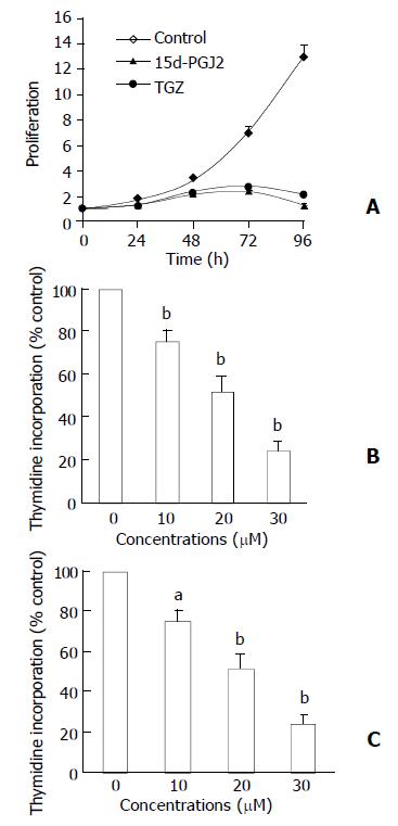
Figure 1 Concentration and time effect of 15d-PGJ2 or TGZ on growth of BEL-7402 cells.
(A) BEL-7402 cells were incubated with 20 µM 15d-PGJ2 or TGZ for 12, 24, 48, 72, 96 h. (B) BEL-7402 cells were incubated with various concentrations of 15d-PGJ2 for 48 h; (C) BEL-7402 cells were incubated with various concentra-tions of TGZ for 48 h. The value was represented as mean ± SEM (n = 3). aP < 0.05 and bP < 0.01 versus corresponding control group.
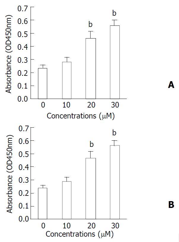
Figure 2 DNA fragmentation by ELISA assay, as measured by absorbance (OD 450 values).
Culture of BEL-7402 cells for 48 h in the presence of 15d-PGJ2/TGZ resulted in dose de-pendent DNA fragmentation. A) 15d-PGJ2; B) TGZ. bP < 0.01 compared to respective control. The value was represented as mean ± SEM (n = 3).
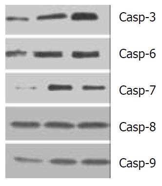
Figure 3 Western blot analysis of the activities of Caspases-3, -6, -7, -8 and –9 in human liver cancer cell line BEL-7402 cells: lane 1: control, lane 2: 30 µM TGZ treated BEL-7402 cells; lane 3: 30 µM 15d-PGJ2 treated BEL-7402 cells.
- Citation: Li MY, Deng H, Zhao JM, Dai D, Tan XY. Peroxisome proliferator-activated receptor gamma ligands inhibit cell growth and induce apoptosis in human liver cancer BEL-7402 cells. World J Gastroenterol 2003; 9(8): 1683-1688
- URL: https://www.wjgnet.com/1007-9327/full/v9/i8/1683.htm
- DOI: https://dx.doi.org/10.3748/wjg.v9.i8.1683


