Copyright
©The Author(s) 2003.
World J Gastroenterol. Jun 15, 2003; 9(6): 1318-1322
Published online Jun 15, 2003. doi: 10.3748/wjg.v9.i6.1318
Published online Jun 15, 2003. doi: 10.3748/wjg.v9.i6.1318
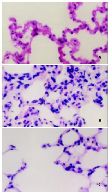
Figure 1 Light microscopic observation on the lung after IIR with pretreatment of AG in rats (HE × 400).
A. The normal lung tissue structure was found in sham group; B. Lung edema, hemorrhage and inflammatory cells sequestration were found in the IR group; C. Decreased morphological changes induced by the intestinal IR were found in the IR + AG group.
Figure 2 Western blotting analysis of iNOS in rat lung after IIR with pretreatment of AG in rats.
1. Sham; 2. IR; 3. IR + AG.
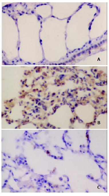
Figure 3 Immunohistochemical analysis of NT in the lung after IIR with pretreatment of AG in rats.
SP stain × 400. A. No positive signal was found in the lung in sham group; B. Intense positive NT staining was found in the IR group; C. Positive NT staining decreased in the IR + AG group.
- Citation: Zhou JL, Jin GH, Yi YL, Zhang JL, Huang XL. Role of nitric oxide and peroxynitrite anion in lung injury induced by intestinal ischemia-reperfusion in rats. World J Gastroenterol 2003; 9(6): 1318-1322
- URL: https://www.wjgnet.com/1007-9327/full/v9/i6/1318.htm
- DOI: https://dx.doi.org/10.3748/wjg.v9.i6.1318


