Copyright
©The Author(s) 2003.
World J Gastroenterol. Jun 15, 2003; 9(6): 1265-1269
Published online Jun 15, 2003. doi: 10.3748/wjg.v9.i6.1265
Published online Jun 15, 2003. doi: 10.3748/wjg.v9.i6.1265
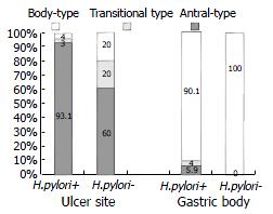
Figure 1 Prevalence of antral-type mucosa at the edge of proximal gastric ulcers and non-ulcerated gastric body in H.
pylori-positive patients (H. pylori+, n = 101) and those without H. pylori infection (H. pylori-, n = 15).
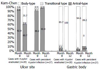
Figure 2 Presence of antral-type mucosa at the edge of proximal gastric ulcers and non-ulcerated gastric body in patients with H.
pylori eradicated (n = 28) and in those with persistent infection (n = 37) before (month 0) and 12 mo after treatment.
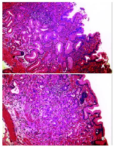
Figure 3 Gastric mucosa at the ulcer edge before and after eradication of H.
pylori infection in the same patient. A, biopsy of the ulcer edge before H. pylori eradication showing antral-type gastric mucosa with severe active chronic inflammation; B, biopsy of the healed ulcer site after H. pylori eradication showing body-type gastric mucosa with presence of parietal and chief cells in the gastric glands and mild residual chronic inflammation. Haematoxylin & eosin (H&E) staining × 250.
-
Citation: Xia HHX, Lam SK, Wong WM, Hu WHC, Lai KC, Wong SH, Leung SY, Yuen ST, Wright NA, Wong BCY. Antralization at the edge of proximal gastric ulcers: Does
Helicobacter pylori infection play a role? World J Gastroenterol 2003; 9(6): 1265-1269 - URL: https://www.wjgnet.com/1007-9327/full/v9/i6/1265.htm
- DOI: https://dx.doi.org/10.3748/wjg.v9.i6.1265


