Copyright
©The Author(s) 2003.
World J Gastroenterol. Jun 15, 2003; 9(6): 1174-1178
Published online Jun 15, 2003. doi: 10.3748/wjg.v9.i6.1174
Published online Jun 15, 2003. doi: 10.3748/wjg.v9.i6.1174
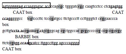
Figure 1 The sequence of ECRG 4 fregment for bisulfite- DHPLC analysis.
The fragment contains 4 cis-acting elements and 16 CpG sites in shadow. The 5'and 3'primers are in the frames of the two ends of the fragment respectively.
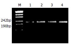
Figure 2 The 1.
5% agarose gel detection of the ssPCR products of ECRG 4. M; pUC19 DNA/Msp I Hap II) Marker. 1, 2, 3, 4; four tissue samples.
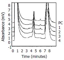
Figure 3 The sizing analysis of ssPCR products of ECRG 4 on DHPLC at 48 °C.
PC was the product from the Sss I treated human placenta genomic DNA. 1, 2, 3, 4; the same samples as in Figure 2.
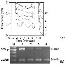
Figure 4 (a) The methylation detection of ssPCR products at 56 °C on DHPLC.
The methylation peak was emphasized by the arrow. PC was the product from the SssI treated human placenta genomic DNA. 1, 2, 3, 4; the same samples as Figure 2. (b) The expression level of ECRG 4 by RT-PCR using the primer set flanking the ORF of the gene in the same samples was detected on DHPLC. The β-Actin gene was amplified as internal control. M; 1 kb DNA Ladder Marker. 1, 2, 3, 4; the same samples as Figure 2.
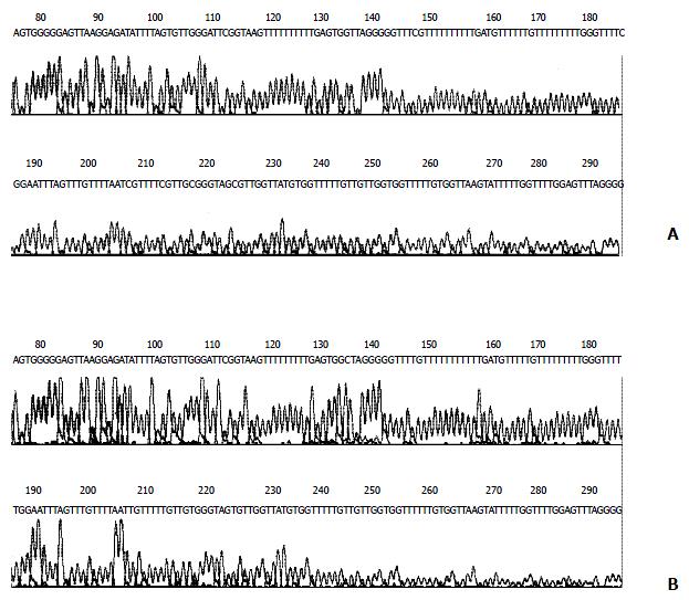
Figure 5 Sequencing of ssPCR products of the ECRG4 gene promoter region.
All cytosines in CpG dinuleotides in the methylated ECRG4 remain as cytosines, indicating methylation (A), while all cytosines in unmethylated ECRG4 have been converted to thymidines, indicating unmethylation (B).
- Citation: Yue CM, Deng DJ, Bi MX, Guo LP, Lu SH. Expression of ECRG4, a novel esophageal cancer-related gene, downregulated by CpG island hypermethylation in human esophageal squamous cell carcinoma. World J Gastroenterol 2003; 9(6): 1174-1178
- URL: https://www.wjgnet.com/1007-9327/full/v9/i6/1174.htm
- DOI: https://dx.doi.org/10.3748/wjg.v9.i6.1174


