Copyright
©The Author(s) 2003.
World J Gastroenterol. Jun 15, 2003; 9(6): 1170-1173
Published online Jun 15, 2003. doi: 10.3748/wjg.v9.i6.1170
Published online Jun 15, 2003. doi: 10.3748/wjg.v9.i6.1170
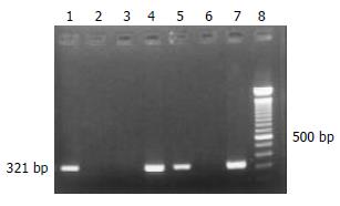
Figure 1 PCR results of Chinese esophageal cancer samples using HPV-16 E6 specific primer.
Lane 1, 2 were the positive and negative control; Lane 4, 5, 7 were the positive samples; Lane 3, 6 were the negative samples; Lane 8 was 100 bp ladder.
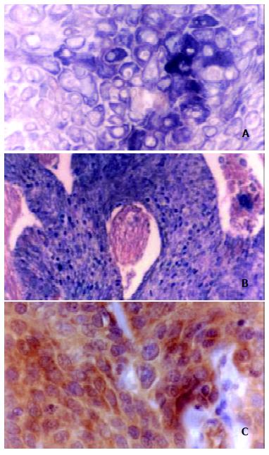
Figure 2 ISH and IHC results of tumor samples targeting HPV-16 E6 gene.
A, the positive purple-blue ISH signal is mainly located in the cytoplasm of esophageal cancer cell. × 200; B, the positive purple-blue ISH signal is mainly located in the nuclear of the carcinoma cell. × 100; C, note the dark-brown IHC signals located mainly in the cytoplasm of cancer cell. SP methods, haematoxylin counterstained × 200.
- Citation: Zhou XB, Guo M, Quan LP, Zhang W, Lu ZM, Wang QH, Ke Y, Xu NZ. Detection of human papillomavirus in Chinese esophageal squamous cell carcinoma and its adjacent normal epithelium. World J Gastroenterol 2003; 9(6): 1170-1173
- URL: https://www.wjgnet.com/1007-9327/full/v9/i6/1170.htm
- DOI: https://dx.doi.org/10.3748/wjg.v9.i6.1170


