Copyright
©The Author(s) 2003.
World J Gastroenterol. Mar 15, 2003; 9(3): 568-571
Published online Mar 15, 2003. doi: 10.3748/wjg.v9.i3.568
Published online Mar 15, 2003. doi: 10.3748/wjg.v9.i3.568
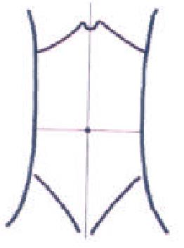
Figure 1 Abridged general view of dog abdomen subregion.
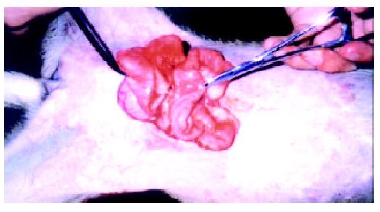
Figure 2 Adhesions between the bowls of animal in control group.
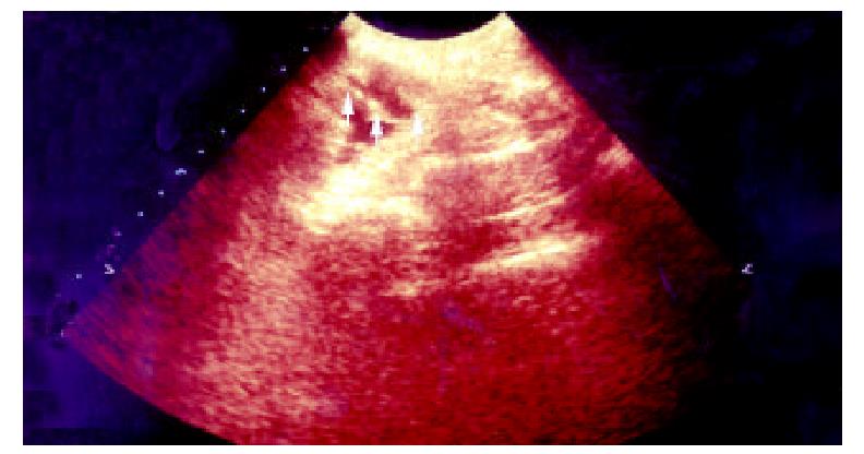
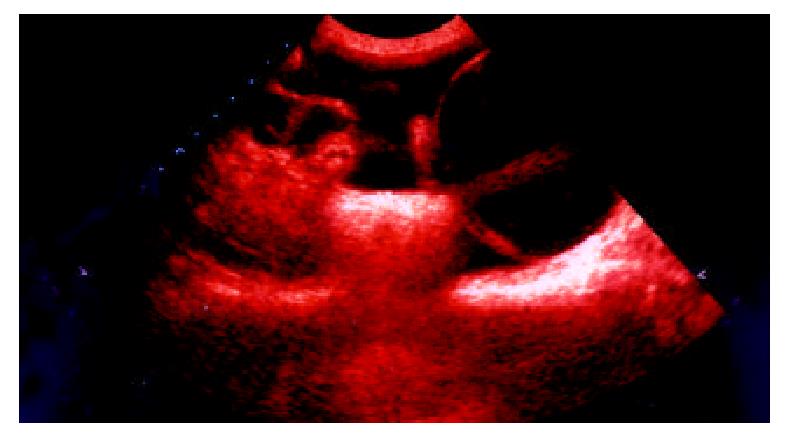
Figure 3 Transabdominal sonogram showed the adhesions between bowls, the adhesion looks like a mouse tail, and the score is 1.
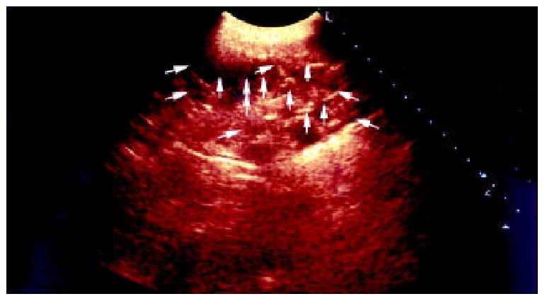
Figure 4 Transabdominal sonogram showed massive agglu-tinating adhesion between bowls, and the score is 3.
Figure 5 Transabdominal sonogram showed the adhesions between bladder and bowls, and the score is 4.
- Citation: Wang XC, Gui CQ, Zheng QS. Combined therapy of allantoin, metronidazole, dexamethasone on the prevention of intra-abdominal adhesion in dogs and its quantitative analysis. World J Gastroenterol 2003; 9(3): 568-571
- URL: https://www.wjgnet.com/1007-9327/full/v9/i3/568.htm
- DOI: https://dx.doi.org/10.3748/wjg.v9.i3.568


