Copyright
©The Author(s) 2003.
World J Gastroenterol. Feb 15, 2003; 9(2): 295-299
Published online Feb 15, 2003. doi: 10.3748/wjg.v9.i2.295
Published online Feb 15, 2003. doi: 10.3748/wjg.v9.i2.295
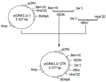
Figure 1 Construction of p/TN.
cDNA coding for hEDN or HBVc was amplified by RT-PCR, ligated together, and then cloned into pcDNA3.1 (-) to form p/TN.
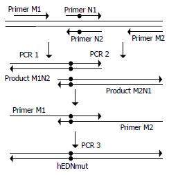
Figure 2 Generation of hEDNmut gene by sequential PCR.
For PCR 1 and 2, pUC18/hEDN was used as template. Primers pair M1 and N2 were used in PCR 1 and N1 and M2 in PCR 2. For PCR 3, the mixture of PCR 1 and 2 product was used as template and M1 and M2 as primers. The black dots in the figure denote the nucleotides introducing the Lys113→Arg mutation which eliminates the ribonuclease activity.
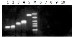
Figure 3 Confirmation of transgenes expression in transfected 2.
2.15 cell line. Forty-eight h after the transfection, total RNA was isolated from transfected 2.2.15 cells and RT-PCR was performed to confirm the expression of transgenes. The RT-PCR products were then electrophoresed in 1.2% agarose gel. Lanes 1-5 represent the RT-PCR results for total RNA isolated from 2.2.15 cells transfected by p/TN, p/TNmut, p/HBVc, p/hEDN, and pcDNA3.1 (-), respectively. Lanes 6-10 represent controls corresponding to lanes 1-5, respectively, in which reverse transcriptase was omitted in RT-PCR. M: DNA Marker (2000, 1000, 750, 500, 250, 100bp from top to bottom).
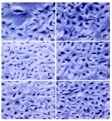
Figure 4 Morphology of 2.
2.15 cells 48 h after transfection. A-F represent 2.2.15 cells transfected by p/TN, p/hEDN, p/HBVc, p/TNmut, pcDNA3.1 (-), or mock transfection, respectively. There were no discernible morphological differences of 2.2.15 cells transfected with p/TN, as compared with the controls.
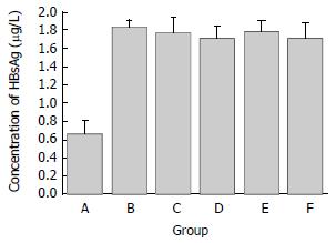
Figure 5 The HBsAg concentration in supernatants of transfected 2.
2.15 cells. Groups A-F represent 2.2.15 cells transfected by p/TN, p/hEDN, p/HBVc, p/TNmut, pcDNA3.1 (-), or mock transfection, respectively. The concentration of HBsAg in the supernatant of 2.2.15 cells transfected with p/TN was decreased by 58% as compared with that of mock transfected 2.2.15 cells.
- Citation: Liu J, Li YH, Xue CF, Ding J, Gong WD, Zhao Y, Huang YX. Targeted ribonuclease can inhibit replication of hepatitis B virus. World J Gastroenterol 2003; 9(2): 295-299
- URL: https://www.wjgnet.com/1007-9327/full/v9/i2/295.htm
- DOI: https://dx.doi.org/10.3748/wjg.v9.i2.295


