Copyright
©The Author(s) 2003.
World J Gastroenterol. Feb 15, 2003; 9(2): 267-270
Published online Feb 15, 2003. doi: 10.3748/wjg.v9.i2.267
Published online Feb 15, 2003. doi: 10.3748/wjg.v9.i2.267
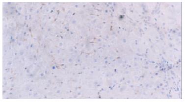
Figure 1 IGF-II expression in the cytoplasm of sinusoidal cells in the paracancerous cirrhotic tissues.
Immunohistochemical staining, × 200.
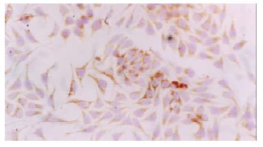
Figure 2 IGF-II expression in the cytoplasm of SMMC7721 cell line.
Immunocytochemical staining, × 200.
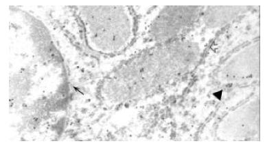
Figure 3 Colloidal gold particles in rough endoplasmic reticulum (↑), mitochondrion (▲), nucleus and paranuclear region (←).
Electron microscopic immunocytochemical staining for IGF-II, × 16000.
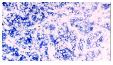
Figure 4 Expression of IGF-II mRNA in the cytoplasm of malignant hepatocytes.
In situ hybridization, × 200.
- Citation: Wang Z, Ruan YB, Guan Y, Liu SH. Expression of IGF-II in early experimental hepatocellular carcinomas and its significance in early diagnosis. World J Gastroenterol 2003; 9(2): 267-270
- URL: https://www.wjgnet.com/1007-9327/full/v9/i2/267.htm
- DOI: https://dx.doi.org/10.3748/wjg.v9.i2.267


