Copyright
©The Author(s) 2003.
World J Gastroenterol. Feb 15, 2003; 9(2): 262-266
Published online Feb 15, 2003. doi: 10.3748/wjg.v9.i2.262
Published online Feb 15, 2003. doi: 10.3748/wjg.v9.i2.262
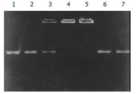
Figure 1 Determination of encapsulation coefficiency.
Lane 1: pure pcDNA3.0/endostatin plasmid, lanes 2-5: TF:liposome:DNA (mg:nmol:mg) at a ratio of 10:1:1, 10:5:1, 10:10:1 and 10:20:1. Lanes 6-7: complexes dissociated with 4 g·L-1 SDS.
Figure 2 Expression of endostatin in vitro.
Lane 1: HUVEC cells transfected with pcDNA3.0/endostatin; lane 2: supernatant of HUVEC cells transfected with pcDNA3.0/endostatin; lane 3: HUVEC cells transfected with pcDNA3.0; lane 4: supernatant of HUVEC cells transfected with pcDNA3.0.
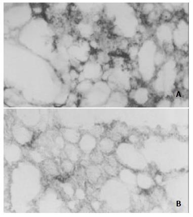
Figure 3 Expression of pcDNA3.
0/endostatin in vivo. The lung tissue sections from mice treated with TF-liposome-pcDNA3.0/endostatin (A) and from mice treated with TF-liposome- pcDNA3.0 (B) were treated with anti-HA antibody.
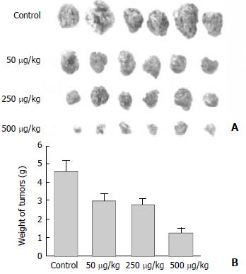
Figure 4 TF-liposome containing endostatin inhibits tumors growth.
A: Tumors excised from mice treated with different doses of TF-liposome-pcDNA3.0/endostatin, or with TF-liposome-pcDNA3.0 (negative control). B: (PDF) Tumor weight of mice treated with TF-liposome-pcDNA3.0/endostatin or TF-liposome-pcDNA3.0 vector. Each bar represents the mean ± SD for six mice.
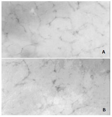
Figure 5 Reduced angiogenesis in TF-liposome-pcDNA3.
0/endostatin treated tumors. Tumor sections from mice treated with TF-liposome-pcDNA3.0 (A) or treated with TF-liposome-pcDNA3.0/endostatin (B) were stained with a CD-31 monoclonal antibody. CD-31-positive cells were brown whereas negative cells colorless in stain.
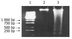
Figure 6 The introduced endostatin gene induces apoptosis in cells of tumor tissues.
DNA was extracted from tumors and DNA fragmentation identified on a 15 g·L-1 agarose gel. Lane 1: the DNA molecular weight marker; lane 2: DNA from mice treated with TF-liposome-pcDNA3.0 vector; lane 3: DNA from mice treated with TF-liposome-pcDNA3.0/endostatin.
- Citation: Li X, Fu GF, Fan YR, Shi CF, Liu XJ, Xu GX, Wang JJ. Potent inhibition of angiogenesis and liver tumor growth by administration of an aerosol containing a transferrin-liposome-endostatin complex. World J Gastroenterol 2003; 9(2): 262-266
- URL: https://www.wjgnet.com/1007-9327/full/v9/i2/262.htm
- DOI: https://dx.doi.org/10.3748/wjg.v9.i2.262


