Copyright
©The Author(s) 2003.
World J Gastroenterol. Dec 15, 2003; 9(12): 2745-2750
Published online Dec 15, 2003. doi: 10.3748/wjg.v9.i12.2745
Published online Dec 15, 2003. doi: 10.3748/wjg.v9.i12.2745
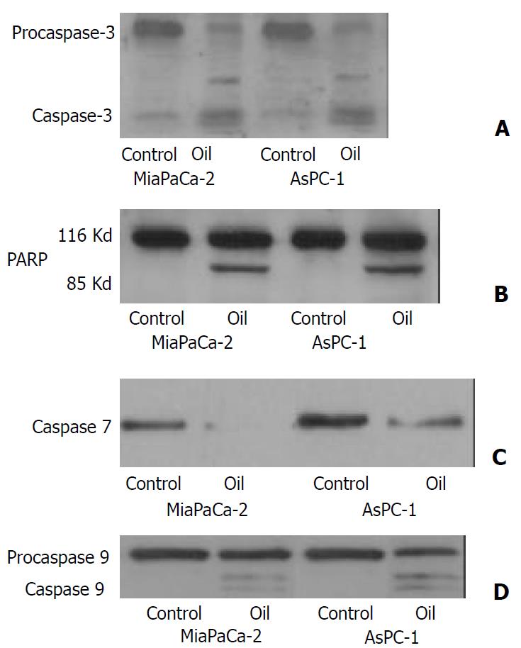
Figure 1 (A, B, C, D) Western blotting of the caspase-3, poly-ADP ribose polymerase (PARP), caspase 7 and caspase 9 in MiaPaCa-2 and AsPC-1 cells treated with 1:32000 oil A for 24 hours.
Proteins in whole cellular lysates were electrophoresed in SDS-PAGE gels and transferred to nitrocellulose membranes. Caspase-3, PARP, caspase 7 and caspase 9 were identified us-ing specific antibodies.
Figure 2 Western blotting of the cytochrome c protein in MiaPaCa-2 and AsPC-1 cells treated with 1:32000 oil A for 24 hours.
Proteins in cytosolic fraction and in mitochondria frac-tion were extracted. Proteins extracted from each sample were electrophoresed on an SDS-PAGE gel and transferred to nitro-cellulose membrane. Cytochrome c was identified using a monoclonal cytochrome c antibody.
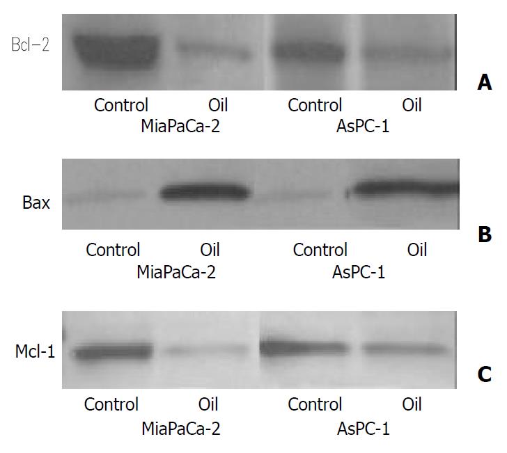
Figure 3 (A, B, C) Western blotting of the Bcl-2, Bax and Mcl-1 proteins in MiaPaCa-2 and AsPC-1 cells treated with 1:32000 oil A for 24 hours.
Whole cellular lysates were electrophore-sed in SDS-PAGE gels and proteins were transferred to nitro-cellulose membranes. Bcl-2, Bax and Mcl-1 were identified us-ing specific antibodies.
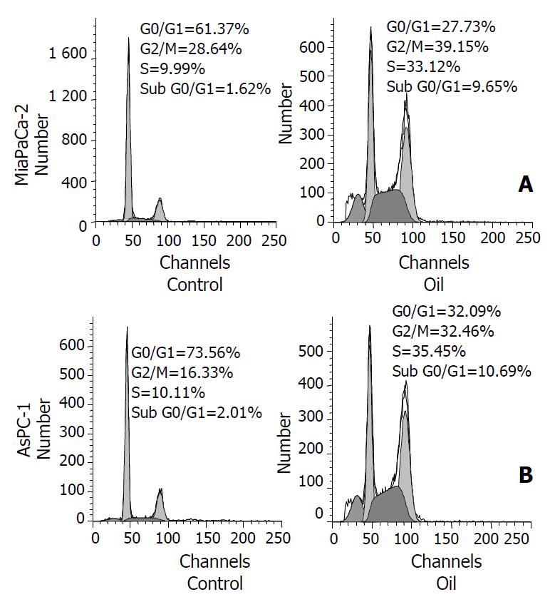
Figure 4 (A, B) Flow-cytometric analysis of cellular DNA con-tent in control and oil A-treated MiaPaCa-2 and AsPC-1 cells, stained with propidium iodide.
The cells were treated with 1:32000 oil A in serum-free conditions for 24 hours. The dis-tribution of cell cycle phases is expressed as% of total cells.
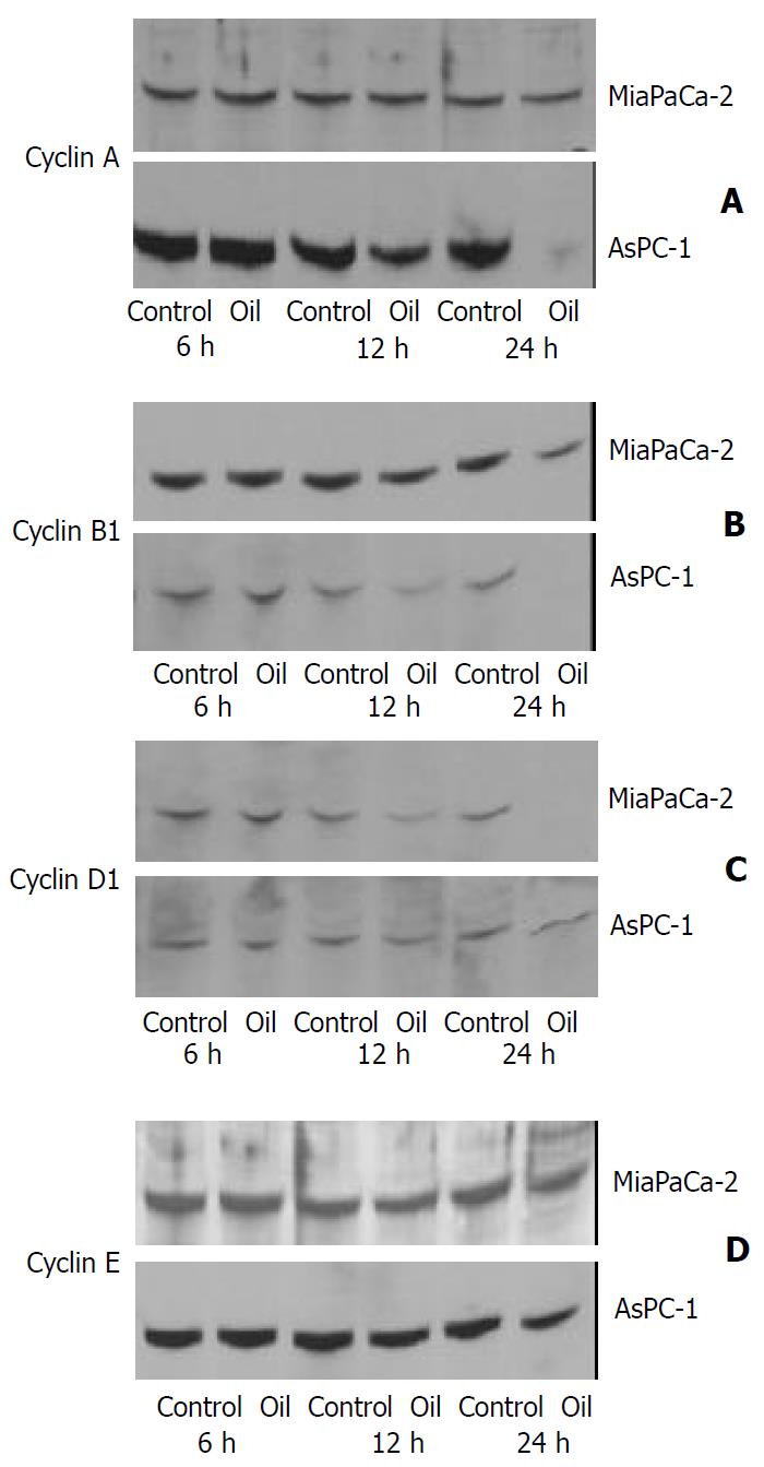
Figure 5 (A, B, C, D) Western blotting of the cyclin A, cyclin B1, cyclin D1 and cyclin E proteins in MiaPaCa-2 and AsPC-1 cells treated with 1:32000 oil A for 24 hours.
Whole cellular lysates were electrophoresed in SDS-PAGE gels and proteins were transferred to nitrocellulose membranes.
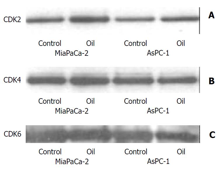
Figure 6 (A, B, C) Western blotting of the CDK2, CDK4 and CDK6 proteins in MiaPaCa-2 and AsPC-1 cells treated with 1:32000 oil A for 24 hours.
Whole cellular lysates were elec-trophoresed in SDS-PAGE gels and proteins were transferred to nitrocellulose membranes.
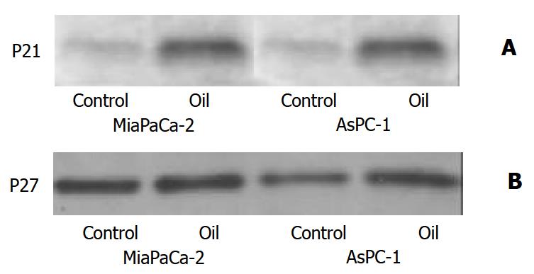
Figure 7 (A, B) Western blotting of the P21 and P27 proteins in MiaPaCa-2 and AsPC-1 cells treated with 1:32000 oil A for 24 hours.
Whole cellular lysates were electrophoresed in SDS-PAGE gels and proteins were transferred to nitrocellulose membranes. P21 and P27 were identified using specific antibodies.
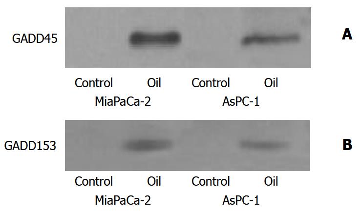
Figure 8 (A, B) Western blotting of GADD45 and GADD153 proteins in MiaPaCa-2 and AsPC-1 cells treated with 1:32000 oil A for 24 hours.
Whole cellular lysates were electrophore-sed in SDS-PAGE gels and proteins were transferred to nitro-cellulose membranes. GADD was identified using specific monoclonal antibodies.
- Citation: Dong ML, Zhu YC, Hopkins JV. Oil A induces apoptosis of pancreatic cancer cells via caspase activation, redistribution of cell cycle and GADD expression. World J Gastroenterol 2003; 9(12): 2745-2750
- URL: https://www.wjgnet.com/1007-9327/full/v9/i12/2745.htm
- DOI: https://dx.doi.org/10.3748/wjg.v9.i12.2745


