Copyright
©The Author(s) 2003.
World J Gastroenterol. Dec 15, 2003; 9(12): 2706-2710
Published online Dec 15, 2003. doi: 10.3748/wjg.v9.i12.2706
Published online Dec 15, 2003. doi: 10.3748/wjg.v9.i12.2706
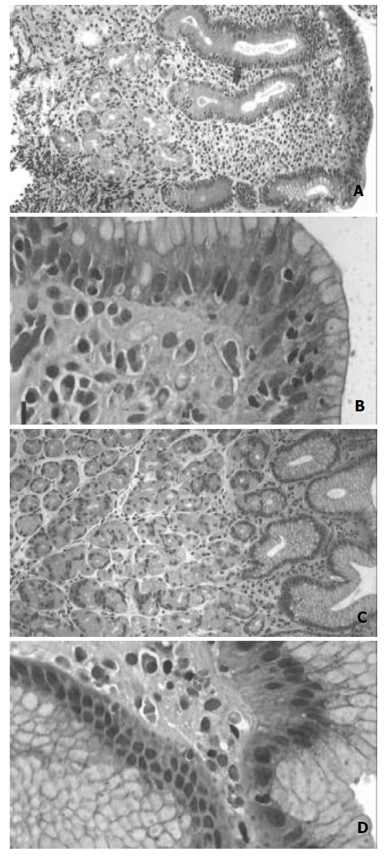
Figure 1 A,B,C,D.
Hematoxylin-Eosin stained sections from gastric body mucosa in a case of lymphocytic gastritis before and after eradication treatment. Before treatment (A,B) there was a sharp increase of intraepithelial lymphocytes, moderate glandular atrophy and foveolar hyperplasia. After treatment (C,D) there were only occasional intraepithelial lymhocytes and no signs of glandular atrophy or foveolar hyperplasia. A,C, Bar=25 μm. B,D, Bar=10 μm.
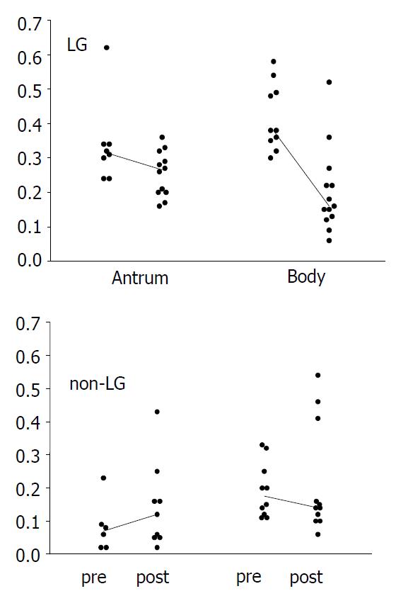
Figure 2 A scatterplot showing epithelial cell proliferation indexes in antral and body mucosa in patients with LG and non-LG H pylori gastritis before (pre) and after (post) treatment.
The median is indicated with a solid line.
-
Citation: Mäkinen JM, Niemelä S, Kerola T, Lehtola J, Karttunen TJ. Epithelial cell proliferation and glandular atrophy in lymphocytic gastritis: Effect of
H pylori treatment. World J Gastroenterol 2003; 9(12): 2706-2710 - URL: https://www.wjgnet.com/1007-9327/full/v9/i12/2706.htm
- DOI: https://dx.doi.org/10.3748/wjg.v9.i12.2706


