Copyright
©The Author(s) 2003.
World J Gastroenterol. Dec 15, 2003; 9(12): 2701-2705
Published online Dec 15, 2003. doi: 10.3748/wjg.v9.i12.2701
Published online Dec 15, 2003. doi: 10.3748/wjg.v9.i12.2701
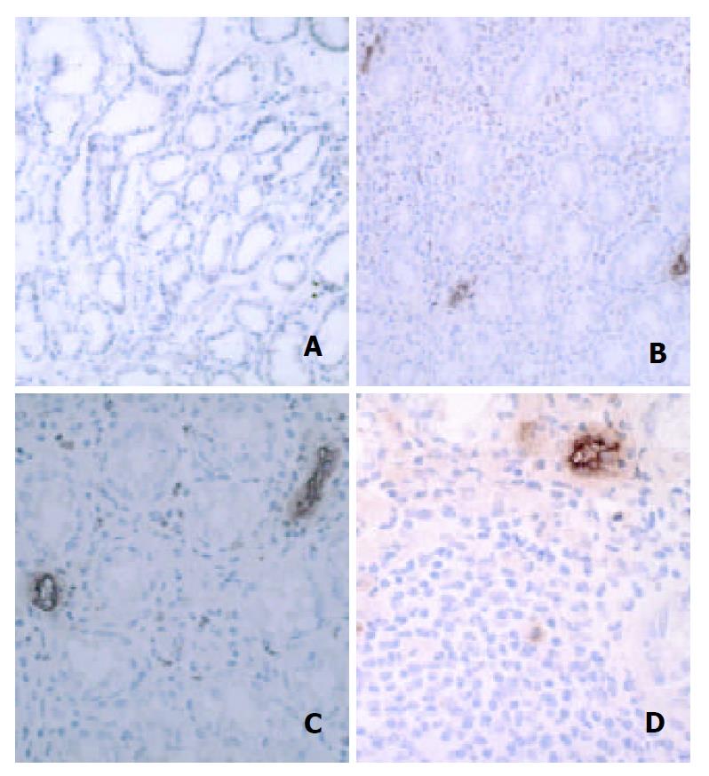
Figure 1 (A): H pylori-negative gastric mucosa showed little immunoreactivity for mucosal addressin cell adhesion mol-ecule 1 (MAdCAM-1).
MAdCAM-1 was expressed on the en-dothelium of numerous vessels within the lamina propria with H pylori infection (B), particularly in association with dense mononuclear infiltration (C). Strong endothelial expres-sion of MAdCAM-1 was localized on high endothelial venule-like vessels adjacent to the lymphoid follicles D.
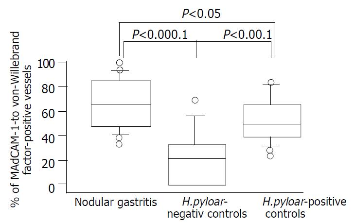
Figure 2 MAdCAM-1 immunoreactivity on mucosal vascula-ture of antral biopsy specimens from patients with nodular gas-tritis and H pylori-positive and –negative controls.
Results were expressed as the percentage of von-Willebrand factor-positive vessels immunoreactive for MAdCAM -1 in serial sections.
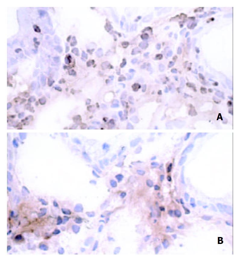
Figure 3 In simultaneous viewing of serial sections, integrin β7-expressing cells (A) consisted of CD-4-positive T lymphocytes (B).
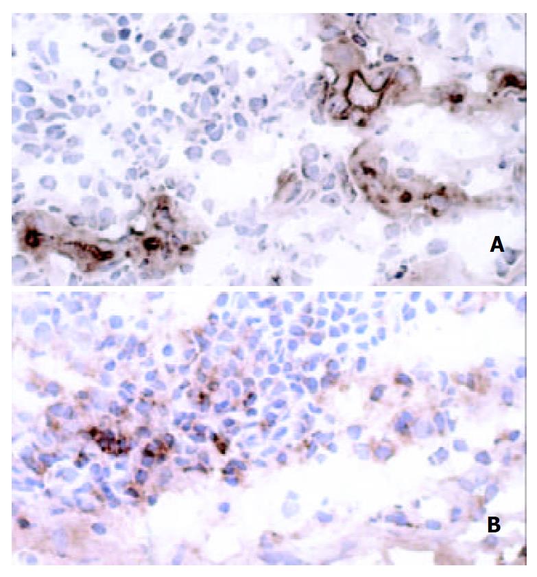
Figure 4 Vessels lined with MAdCAM-1-positive endothelium (A) were associated with infiltration of lymphocytes immu-noreactive for integrin β7 (B).
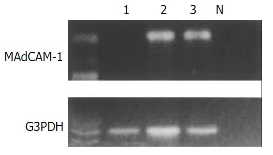
Figure 5 MAdCAM-1 and glyceraldehyde-3-phosphate dehy-drogenase (G3PDH) mRNA transcripts were detected as 387 and 983 base pair-bands with reverse transcriptase-polymerase chain reaction, respectively.
Lane N: negative control, lane 1: H pylori-negative control, lane 2: H pylori-positive control, lane 3: patients with nodular gastritis.
- Citation: Ohara H, Isomoto H, Wen CY, Ejima C, Murata M, Miyazaki M, Takeshima F, Mizuta Y, Murata I, Koji T, Nagura H, Kohno S. Expression of mucosal addressin cell adhesion molecule 1 on vessel endothelium of gastric mucosa in patients with nodular gastritis. World J Gastroenterol 2003; 9(12): 2701-2705
- URL: https://www.wjgnet.com/1007-9327/full/v9/i12/2701.htm
- DOI: https://dx.doi.org/10.3748/wjg.v9.i12.2701


