Copyright
©The Author(s) 2003.
World J Gastroenterol. Oct 15, 2003; 9(10): 2379-2381
Published online Oct 15, 2003. doi: 10.3748/wjg.v9.i10.2379
Published online Oct 15, 2003. doi: 10.3748/wjg.v9.i10.2379
Figure 1 A: Computed tomography revealing a large tumor in the left lobe.
B: Celiac angiography during the arterial phase, showing a large hypervascular lesion.
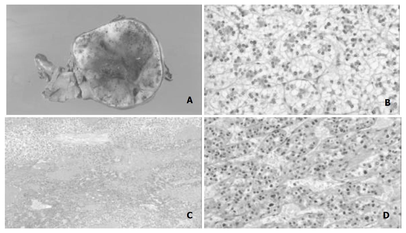
Figure 2 A: Macroscopic appearance of resected tumor.
The tumor was incompletely encapsulated by thin fibrous tissues, and its cut surface was tan-yellowish. Hemorrhage was observed inside part of the tumor. B: Microscopic appearance of tumor. Tumor cells were relatively uniform and had clear eosinophilic cytoplasm with small round nuclei. C: Trabecular structures were spo-radically encountered in association with hemorrhage. D: The tumor cells were arranged predominantly in a thin trabecular pattern with moderate nuclear atypia.
Figure 3 Immunohistochemistry.
A: PIVKA-II. B: CD34. PIVKA-II was positive in the tumor cells with or without nuclear atypism and trabecular structure (A). CD34 was expressed diffusely in the sinusoidal endothelial cells (B).
- Citation: Ito M, Sasaki M, Wen CY, Nakashima M, Ueki T, Ishibashi H, Yano M, Kage M, Kojiro M. Liver cell adenoma with malignant transformation: A case report. World J Gastroenterol 2003; 9(10): 2379-2381
- URL: https://www.wjgnet.com/1007-9327/full/v9/i10/2379.htm
- DOI: https://dx.doi.org/10.3748/wjg.v9.i10.2379


