Copyright
©The Author(s) 2003.
World J Gastroenterol. Oct 15, 2003; 9(10): 2174-2177
Published online Oct 15, 2003. doi: 10.3748/wjg.v9.i10.2174
Published online Oct 15, 2003. doi: 10.3748/wjg.v9.i10.2174
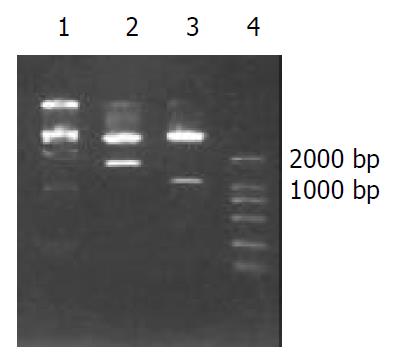
Figure 1 Restricted endonuclease analysis of recombinant plas-mid PCI-asHAb18G.
1: DNA marker DL15000, 2: PCI-asHAb18G/Xho I + Xba I, 3: PCI-asHAb18G/SmaI, 4: DNA marker DL2000.
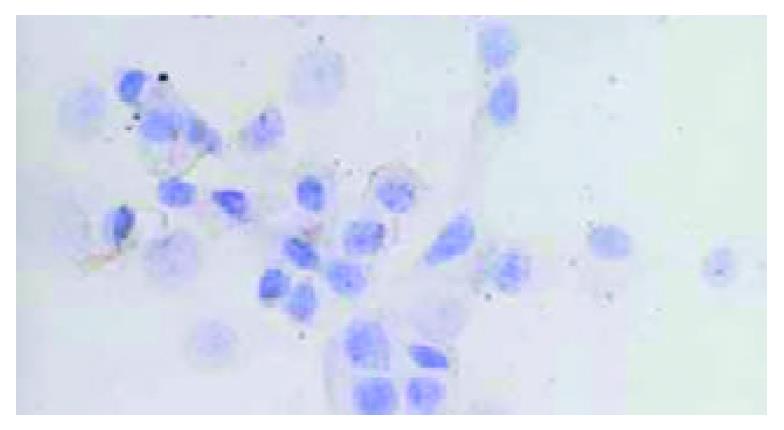
Figure 2 Negative staining of HAb18G/CD147 on membrane of HHCC/asHAb18G SP × 400.
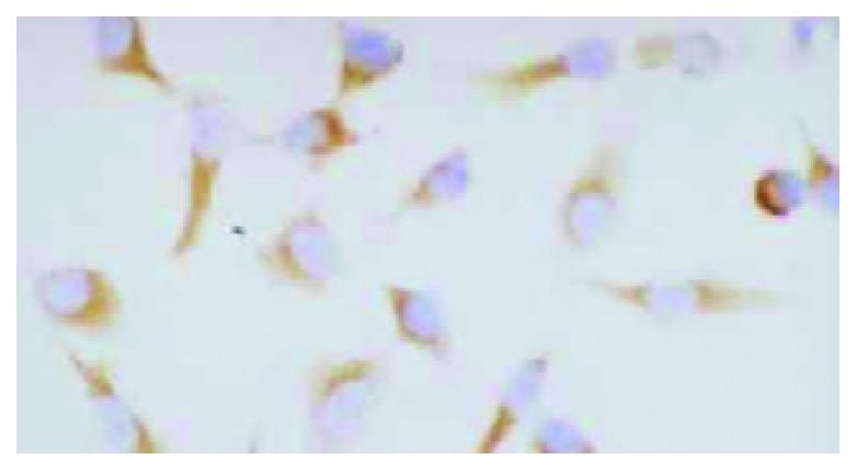
Figure 3 Positive staining of HAb18G/CD147 on membrane of HHCC/neo SP × 400.
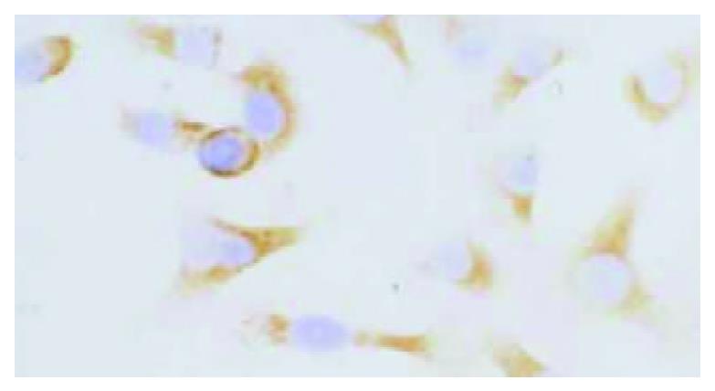
Figure 4 Positive staining of HAb18G/CD147 on membrane of HHCC SP × 400.
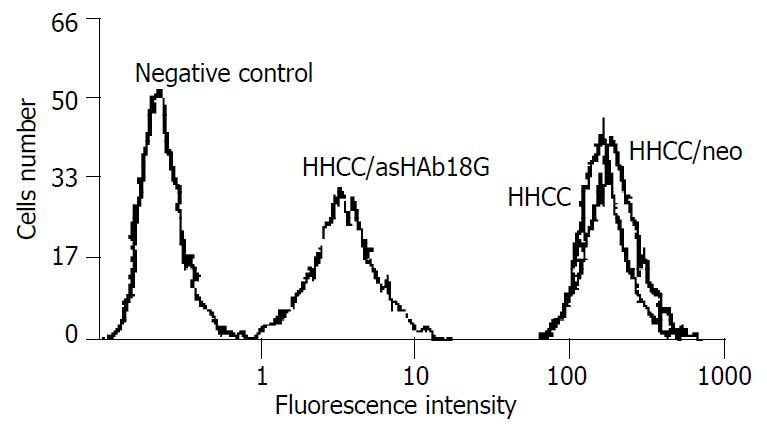
Figure 5 FACS analysis of expressions of HAb18G/CD147 in four kinds of cells.
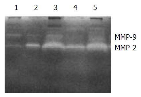
Figure 6 Gelatin zymography of secretions of MMP-2 and MMP-9 in five groups of cells.
1: fb, 2: HHCC, 3: HHCC/neo + fb, 4: HHCC/asHAb18G + fb, 5: HHCC + fb.
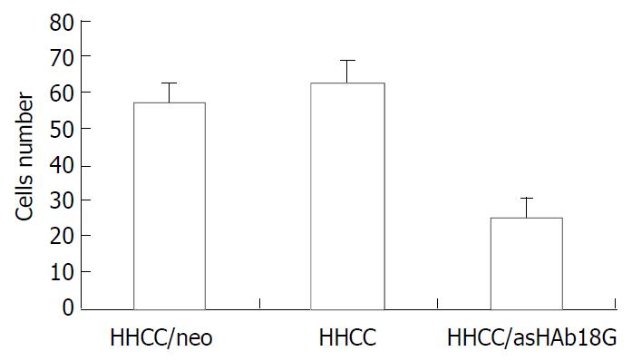
Figure 7 Inhibitory effects of antisense RNA on invasion of HHCC/asHAb18G.
-
Citation: Li Y, Shang P, Qian AR, Wang L, Yang Y, Chen ZN. Inhibitory effects of antisense RNA of HAb18G/CD147 on invasion of hepatocellular carcinoma cells
in vitro . World J Gastroenterol 2003; 9(10): 2174-2177 - URL: https://www.wjgnet.com/1007-9327/full/v9/i10/2174.htm
- DOI: https://dx.doi.org/10.3748/wjg.v9.i10.2174


