Copyright
©The Author(s) 2002.
World J Gastroenterol. Dec 15, 2002; 8(6): 966-970
Published online Dec 15, 2002. doi: 10.3748/wjg.v8.i6.966
Published online Dec 15, 2002. doi: 10.3748/wjg.v8.i6.966
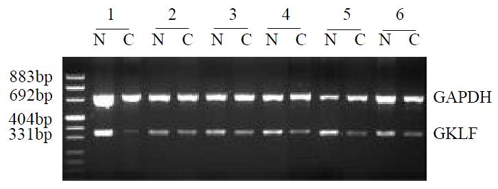
Figure 1 Semi-quantitative RT-PCR of GKLF in esophageal squamous cancer patients.
RNA from specimens of normal appearing mucosa (N) and cancer (C) were extracted and GKLF as well as GAPDH were amplified. This figure showed the rep-resentative results from several individual patients.
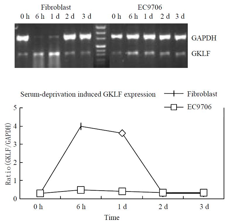
Figure 2 Serum deprivation induced GKLF expression.
Both human primary cultured fibroblast and an esophageal squa-mous cancer cell line EC9706 was underwent serum deprivation. GKLF expression was measured semi-quantitively by RT-PCR. The magnitude of GKLF expression was calcu-lated as ratio of GKLF to GAPDH.
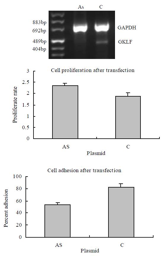
Figure 3 Cell growth and adhesion in GKLF transfected EC9706 cells.
The cells were transiently transfected with antisense GKLF expression plasmid (AS) and pCDNA3.1 as control (C). The GKLF expression levels detected by RT-PCR were shown in the upper panel. The cell growth rates were shown in the middle panel; each data point represents mean ± S.E.M. of 7 repeats; there was a significant difference between AS and C (P < 0.05). The cell adhesions were shown in the lower panel; each data point represents mean ± S.E.M. of 4 repeats; AS had significant difference compared to C (P < 0.05).
- Citation: Wang N, Liu ZH, Ding F, Wang XQ, Zhou CN, Wu M. Down-regulation of gut-enriched Krüppel-like factor expression in esophageal cancer. World J Gastroenterol 2002; 8(6): 966-970
- URL: https://www.wjgnet.com/1007-9327/full/v8/i6/966.htm
- DOI: https://dx.doi.org/10.3748/wjg.v8.i6.966


