Copyright
©The Author(s) 2002.
World J Gastroenterol. Aug 15, 2002; 8(4): 654-657
Published online Aug 15, 2002. doi: 10.3748/wjg.v8.i4.654
Published online Aug 15, 2002. doi: 10.3748/wjg.v8.i4.654
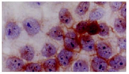
Figure 1 Positive staining of HLA-I antigens at QGY-7703 cell membrane.
LSAB × 350
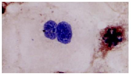
Figure 2 Positive staining of HLA-I antigens in cytoplasm of dividing Alexander cells•LSAB × 350
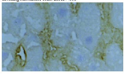
Figure 3 Negative staining of histologically normal liver cells and intensive positive staining within sinusoids.
LSAB × 100.
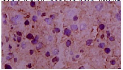
Figure 4 Positive staining of hepatocelluar carcinoma cells LSAB × 100.
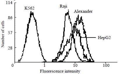
Figure 5 Flow cytometric histogram comparison of HLA-I antigens among K562, Raji, Alexander, HepG2 cell lines.
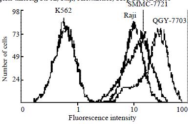
Figure 6 Flow cytometric histogram comparison of HLA-I antigens among K562, Raji, SMMC-7721, QGY-7703 cell lines.
- Citation: Huang J, Cai MY, Wei DP. HLA class I expression in primary hepatocellular carcinoma. World J Gastroenterol 2002; 8(4): 654-657
- URL: https://www.wjgnet.com/1007-9327/full/v8/i4/654.htm
- DOI: https://dx.doi.org/10.3748/wjg.v8.i4.654


