Copyright
©The Author(s) 2002.
World J Gastroenterol. Jun 15, 2002; 8(3): 537-539
Published online Jun 15, 2002. doi: 10.3748/wjg.v8.i3.537
Published online Jun 15, 2002. doi: 10.3748/wjg.v8.i3.537
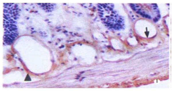
Figure 1 Immunohistochemical stain of eNOS in jejunal submucosa, showing the positive endothelium (↑) and microvascular smooth muscle (▲).
× 170
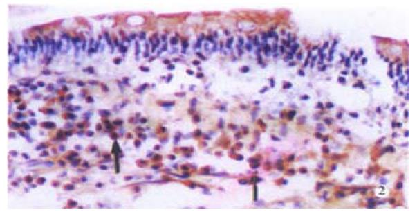
Figure 2 Immunohistochemical stain of eNOS in jejunal villi, showing the positive cell in the proper layer (↑).
× 170
Figure 3 Immunofluorescence histochemical double-stain in jejunum observed under a confocal laser scanning microscope, (a) showing the positive substances of nNOS were distributed mainly in myenteric plexus.
× 350 (b) and (c) showing the nNOS-positive cell (▲), the eNOS-positive cell (★) and double-stained cell (↑) in the proper layer of villi. × 250
- Citation: Chen YM, Qian ZM, Zhang J, Chang YZ, Duan XL. Distribution of constitutive nitric oxide synthase in the jejunum of adult rat. World J Gastroenterol 2002; 8(3): 537-539
- URL: https://www.wjgnet.com/1007-9327/full/v8/i3/537.htm
- DOI: https://dx.doi.org/10.3748/wjg.v8.i3.537


