Copyright
©The Author(s) 2002.
World J Gastroenterol. Feb 15, 2002; 8(1): 36-39
Published online Feb 15, 2002. doi: 10.3748/wjg.v8.i1.36
Published online Feb 15, 2002. doi: 10.3748/wjg.v8.i1.36
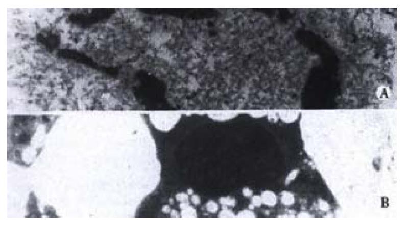
Figure 1 Apoptotic cells in EC/CUHK1 and SGC7901 with the condensa-tion and margination of chromatin, and nuclear breakage EM × 6000
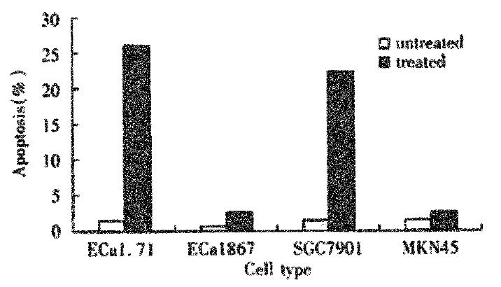
Figure 2 Flow cytometry with PI staining: apoptosis proportions in EC/CUHK1, EC1867, SGC7901 and MKN45
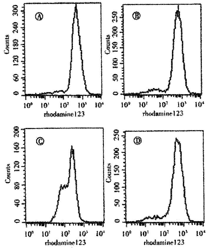
Figure 3 Flow cytometry displaying the inherent ROS level of cells.
A:MKN45; B:SGC7901; C:EC1867; D:EC/CUHK1
- Citation: Gao F, Yi J, Shi GY, Li H, Shi XG, Tang XM. The sensitivity of digestive tract tumor cells to As2O3 is associated with the inherent cellular level of reactive oxygen species. World J Gastroenterol 2002; 8(1): 36-39
- URL: https://www.wjgnet.com/1007-9327/full/v8/i1/36.htm
- DOI: https://dx.doi.org/10.3748/wjg.v8.i1.36


