Copyright
©The Author(s) 2002.
World J Gastroenterol. Feb 15, 2002; 8(1): 26-30
Published online Feb 15, 2002. doi: 10.3748/wjg.v8.i1.26
Published online Feb 15, 2002. doi: 10.3748/wjg.v8.i1.26
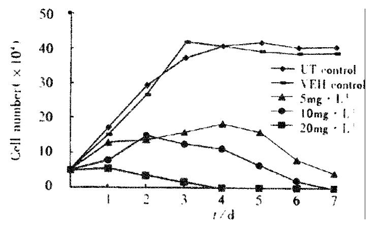
Figure 1 Growth curve of SGC-7901 cell treated with VES.
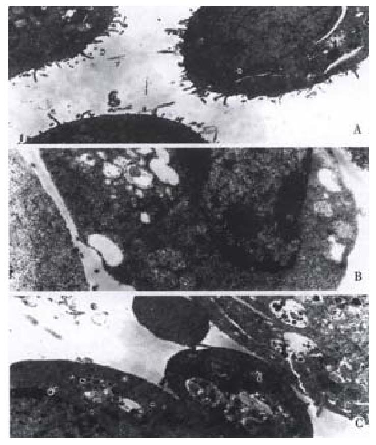
Figure 2 VES-induced apoptosis in SGC-7901 cells with transmission elec-tron microscope.
A: Normal cell; B: Apoptotic cell with chromatin condensation, chromatin crescent formation/margination; C: Cell with apoptotic body.
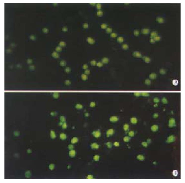
Figure 3 VES-induced apoptosis in SGC-7901 cells with DAPI staining UT control; VES at 20 mg·L-1.
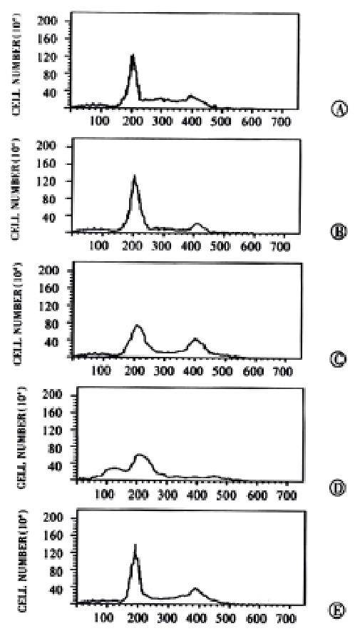
Figure 4 VES-induced apoptosis in SGC-7901 cells with flow cytometry.
A: UT control; B: VEH control; C: VES at 5 mg·L-1; D: VES at 10 mg·L-1; E: VES at 20 mg·L-1.
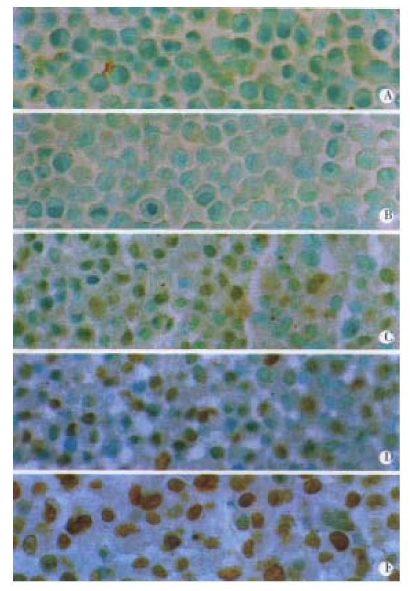
Figure 5 VES-induced apoptosis by TUNEL assay.
A: UT control; B: VEH control; C: VES at 5 mg·L-1; D: VES at 10 mg·L-1; E: VES at 20 mg·L-1.
- Citation: Wu K, Zhao Y, Liu BH, Li Y, Liu F, Guo J, Yu WP. RRR-α-tocopheryl succinate inhibits human gastric cancer SGC-7901 cell growth by inducing apoptosis and DNA synthesis arrest. World J Gastroenterol 2002; 8(1): 26-30
- URL: https://www.wjgnet.com/1007-9327/full/v8/i1/26.htm
- DOI: https://dx.doi.org/10.3748/wjg.v8.i1.26


