Copyright
©The Author(s) 2002.
World J Gastroenterol. Feb 15, 2002; 8(1): 119-123
Published online Feb 15, 2002. doi: 10.3748/wjg.v8.i1.119
Published online Feb 15, 2002. doi: 10.3748/wjg.v8.i1.119
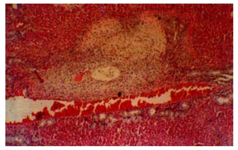
Figure 1 Liver necrosis after HAE in rats.
The necrotic area is seen near the portal triad. HE × 100
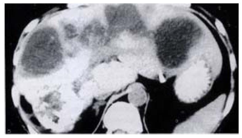
Figure 2 Biliary abscess of liver after HAE.
CT shows multi-abscess along portal tract.
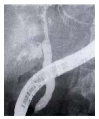
Figure 3 Intrahepatic bile duct stricture after HAE.
ERCP shows biliary stricture.
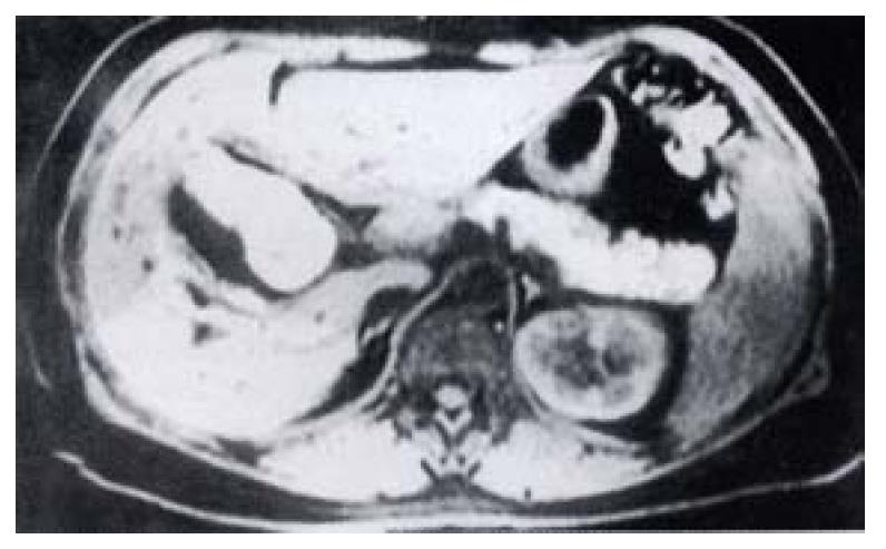
Figure 4 MRI showed fluid around the gallbladder.
- Citation: Huang XQ, Huang ZQ, Duan WD, Zhou NX, Feng YQ. Severe biliary complications after hepatic artery embolization. World J Gastroenterol 2002; 8(1): 119-123
- URL: https://www.wjgnet.com/1007-9327/full/v8/i1/119.htm
- DOI: https://dx.doi.org/10.3748/wjg.v8.i1.119


