Copyright
©The Author(s) 2001.
World J Gastroenterol. Dec 15, 2001; 7(6): 860-863
Published online Dec 15, 2001. doi: 10.3748/wjg.v7.i6.860
Published online Dec 15, 2001. doi: 10.3748/wjg.v7.i6.860
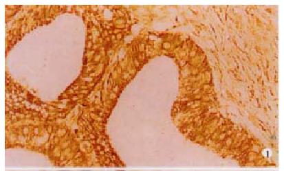
Figure 1 FasL positive in human hilar cholangiocarcinom as (brown).
SABC × 200
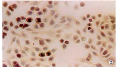
Figure 2 Expression of FasL in cholangiocarcinoma cell line.
QBC939 × 200
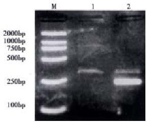
Figure 3 Expression of FasL mRNA in human cholangiocarcinoma cells QBC939.
M: DL 2000 Marker; 1: FasL; 2: FasL+β-actin
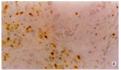
Figure 4 CD45 positive cells (brown) of lymphoid morphology adjacent to carcinoma.
× 200
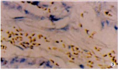
Figure 5 Positive apoptotic TUNEL stainig in situ (brown) with apoptotic morphology.
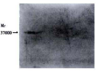
Figure 6 Western blotting of FasL protein with mAb from QBC939 cell cultures clone 33 from QBC939 cell cultures
- Citation: Li ZY, Zou SQ. Fas counterattack in cholangiocarcinoma: A mechanism for immune evasion in human hilar cholangiocarcinomas. World J Gastroenterol 2001; 7(6): 860-863
- URL: https://www.wjgnet.com/1007-9327/full/v7/i6/860.htm
- DOI: https://dx.doi.org/10.3748/wjg.v7.i6.860


