Copyright
©The Author(s) 2001.
World J Gastroenterol. Dec 15, 2001; 7(6): 841-845
Published online Dec 15, 2001. doi: 10.3748/wjg.v7.i6.841
Published online Dec 15, 2001. doi: 10.3748/wjg.v7.i6.841
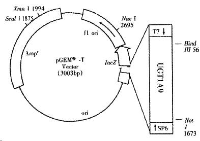
Figure 1 Scheme of pGEM-UGT1A9.
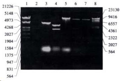
Figure 2 Electrophoresis identification of recombinants constructed.
Lanes 1: Marker (λ/EcoR I and Hind III); 2: PCR product of UGT1A9 from Chinese human liver; 3: recombinant of pGEM-UGT1A9; 4: recombinant of pGEM-UGT1A9 cleaved by Hind III and Not I; 5: recombinant of pREP9-UGT1A9; 6: recombinant of pREP9-UGT1A9 cleaved by Hind III and Not I; 7: pREP9 expression vector; 8: Marker (λ/Hind III).
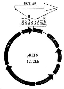
Figure 3 Scheme of pREP9-UGT1A9.
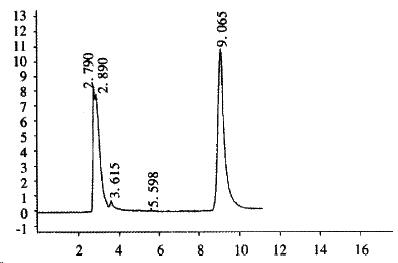
Figure 4 Chromatogram of propranolol after incubation with S9 prepared from CHL-UGT1A9 cell.
A Shim-pack CLC-ODS column (15 cm ± 0.6 cm i.d.) was used. The mobile phase was constituted with ammonium acetate buffer-methanol-acetonitrile (2:2:1) and 1.4 mL•L¯¹ triethylamine with the flow rate at 1.0 mL•min-1. Propranolol was monitored at 290 nm. propranolol: tR = 9.065 min.
- Citation: Li X, Yu YN, Zhu GJ, Qian YL. Cloning of UGT1A9 cDNA from liver tissues and its expression in CHL cells. World J Gastroenterol 2001; 7(6): 841-845
- URL: https://www.wjgnet.com/1007-9327/full/v7/i6/841.htm
- DOI: https://dx.doi.org/10.3748/wjg.v7.i6.841


