Copyright
©The Author(s) 2001.
World J Gastroenterol. Aug 15, 2001; 7(4): 542-546
Published online Aug 15, 2001. doi: 10.3748/wjg.v7.i4.542
Published online Aug 15, 2001. doi: 10.3748/wjg.v7.i4.542
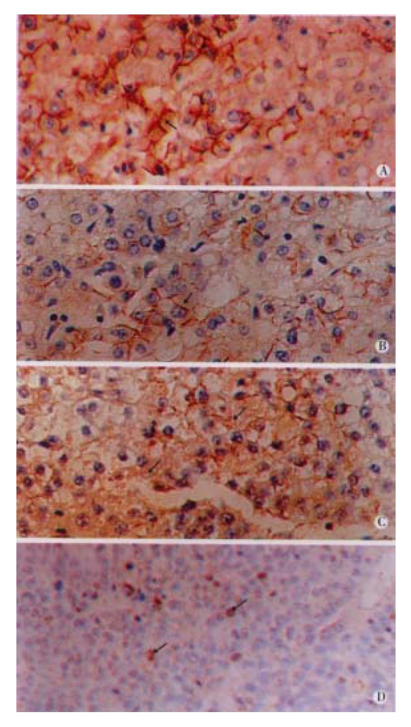
Figure 1 Immunohistochemistry of β-catenin.
A: In normal liver tissue, the staining was mainly positive on the cellular membrane (arrowpoint), with very weak cytoplasmic staining. × 200 B: Para-cancerous cirrhotic liver tissue showed membrane staining (arrowpoint) like normal liver tissue. C,D: For HCC, cytoplasmic and nuclear staining was dominant (arrowpoints), whereas membrane staining was rare. × 200
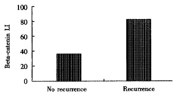
Figure 2 Labeling index (LI) of β-catenin.
Recurrent patient (n = 15) was much higher than that of non-recurrent patient (n = 19) (84.9 ± 17.4) vs (39.1 ± 14.3).
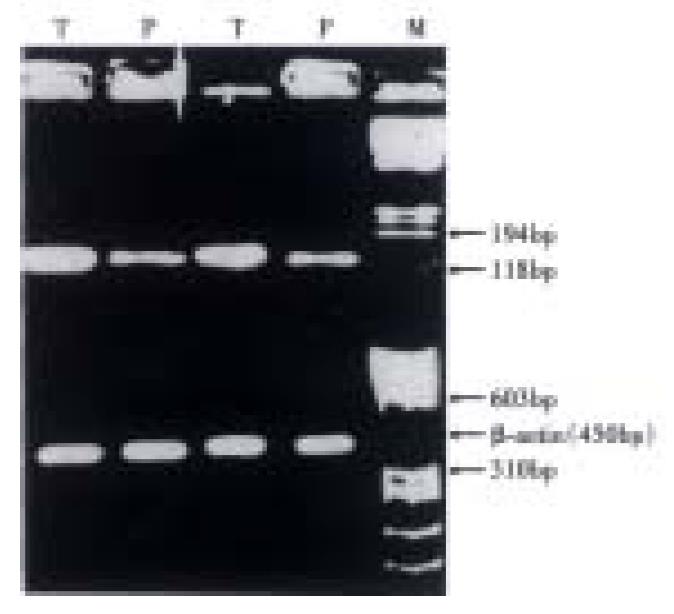
Figure 3 β-catenin mRNA expression index (EI).
HCC was higher vs para-cancerous tissue (P < 0.05). P: para-cancerous tissue; T: HCC; M: nucleic acid molecular mass marker.
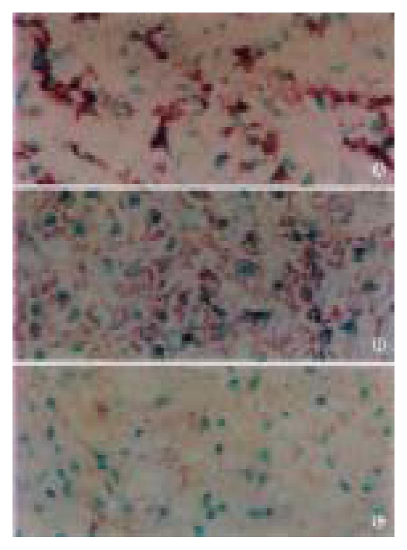
Figure 4 In situ hybridization of β-catenin gene mRNA.
Stronger in HCC (A) vs para-cancerous cirrhotic liver tissue and (B) normal liver tissue (C).
- Citation: Cui J, Zhou XD, Liu YK, Tang ZY, Zile MH. Abnormal β-catenin gene expression with invasiveness of primary hepatocellular carcinoma in China. World J Gastroenterol 2001; 7(4): 542-546
- URL: https://www.wjgnet.com/1007-9327/full/v7/i4/542.htm
- DOI: https://dx.doi.org/10.3748/wjg.v7.i4.542


