Copyright
©The Author(s) 2001.
World J Gastroenterol. Aug 15, 2001; 7(4): 522-526
Published online Aug 15, 2001. doi: 10.3748/wjg.v7.i4.522
Published online Aug 15, 2001. doi: 10.3748/wjg.v7.i4.522
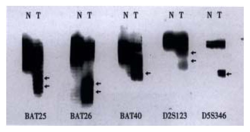
Figure 1 MSI in gastric cancer using 5 microsatellite loci (BAT-25, BAT-26, BAT40, D2S123, and D5S346).
Arrows indicate variant conformers. (N: normal DNA pattern; T: tumor specimens containing variant conformers representing MIS)
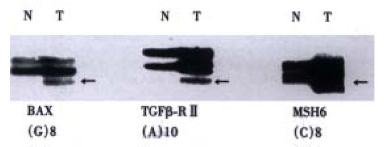
Figure 2 Frameshift mutations of hMSH6 TGF-betaRII, and BAX genes in gastric cancers.
Arrows indicate conformational variants associated with frameshift mutations. (N: normal DNA; T: tumor DNA)
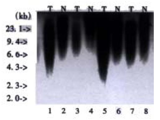
Figure 3 Southern blot analysis of telomere repeat arrays in DNA samples of patients with gastric cancer.
DNA was digested with-Hin fI and hybridized with α-32P-APT end-labeled (TTAGGG)4 probe.T = Tumour; N = Corresponding normal tissues.
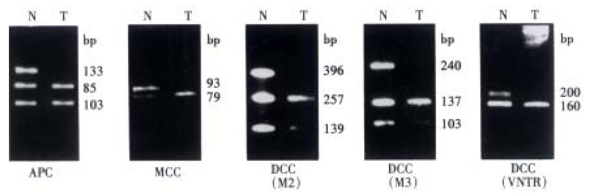
Figure 4 Representative LOH analysis of APC, MCC and DCC genes.
(APC: Rsa1 RFLP in APC exon 11, loss of 133 bp allele is seen in tumor DNA, MCC: A 14 bp insertion/deletion polymorphism in MCC exon 10 gives rise to a 93 or 79 allele. Loss of the 93 bp allele is seen in the tumor. DCC: Losses of 396 bp, 240 bp and 200 bp alleles are seen at M2, M3 and VNTR polymorphic sequences in the tumor DNA)
- Citation: Fang DC, Yang SM, Zhou XD, Wang DX, Luo YH. Telomere erosion is independent of microsatellite instability but related to loss of heterozygosity in gastric cancer. World J Gastroenterol 2001; 7(4): 522-526
- URL: https://www.wjgnet.com/1007-9327/full/v7/i4/522.htm
- DOI: https://dx.doi.org/10.3748/wjg.v7.i4.522


