Copyright
©The Author(s) 2001.
World J Gastroenterol. Jun 15, 2001; 7(3): 363-369
Published online Jun 15, 2001. doi: 10.3748/wjg.v7.i3.363
Published online Jun 15, 2001. doi: 10.3748/wjg.v7.i3.363
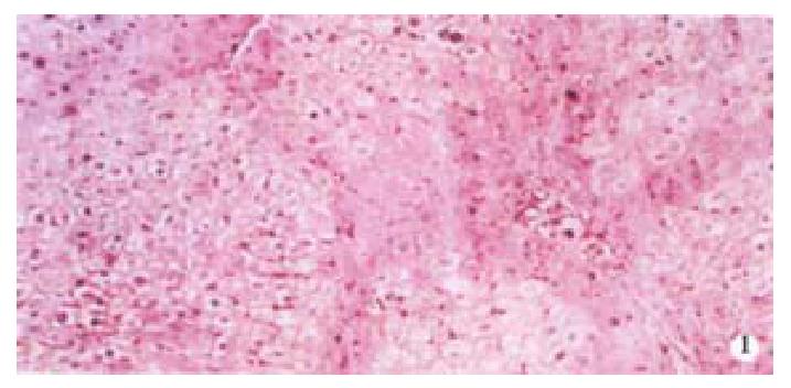
Figure 1 VG staining of rat liver (pseudolobuli were formed).
× 100
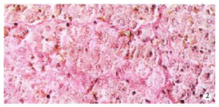
Figure 2 Masson staining of rat liver.
× 200
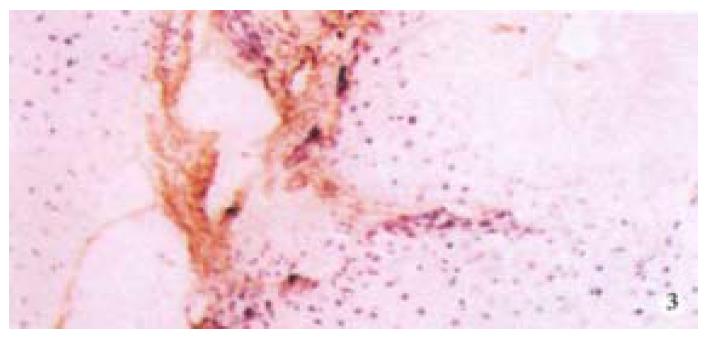
Figure 3 Collagen III in rat liver (immunohisochemistry).
× 200
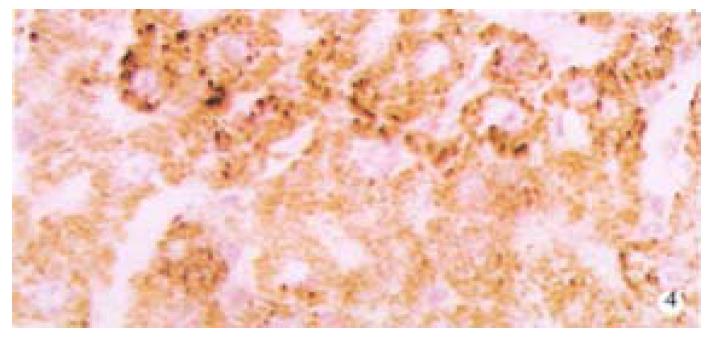
Figure 4 Protein expression of TIMP-1 in rat liver (immunohisochemistry).
× 200
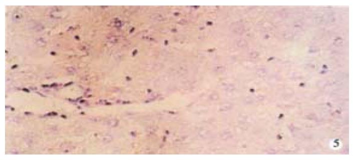
Figure 5 TIMP-1 protein expression of in rat liver after treated with asON.
× 200
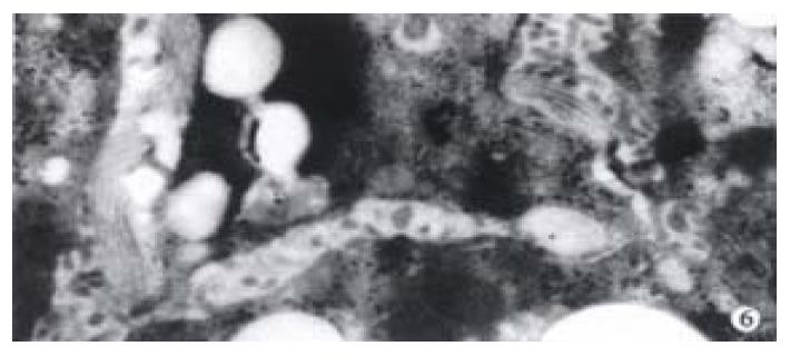
Figure 6 Activated HSC and lots of collagen fibers around HSC in the rat model of hepatic fibrosis EM 10K.
- Citation: Nie QH, Cheng YQ, Xie YM, Zhou YX, Cao YZ. Inhibiting effect of antisense oligonucleotides phosphorthioate on gene expression of TIMP-1 in rat liver fibrosis. World J Gastroenterol 2001; 7(3): 363-369
- URL: https://www.wjgnet.com/1007-9327/full/v7/i3/363.htm
- DOI: https://dx.doi.org/10.3748/wjg.v7.i3.363


