Copyright
©The Author(s) 2001.
World J Gastroenterol. Jun 15, 2001; 7(3): 335-339
Published online Jun 15, 2001. doi: 10.3748/wjg.v7.i3.335
Published online Jun 15, 2001. doi: 10.3748/wjg.v7.i3.335
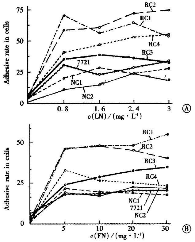
Figure 1 Attachment of different cell clones to LN or FN (-x±sx).
A: To increasing concentrations (0, 0.8, 1.6, 2.4 and 3 mg·L-1) of LN B: To increasing concentrations (0, 5, 10, 20 and 30 mg·L-1) of FN
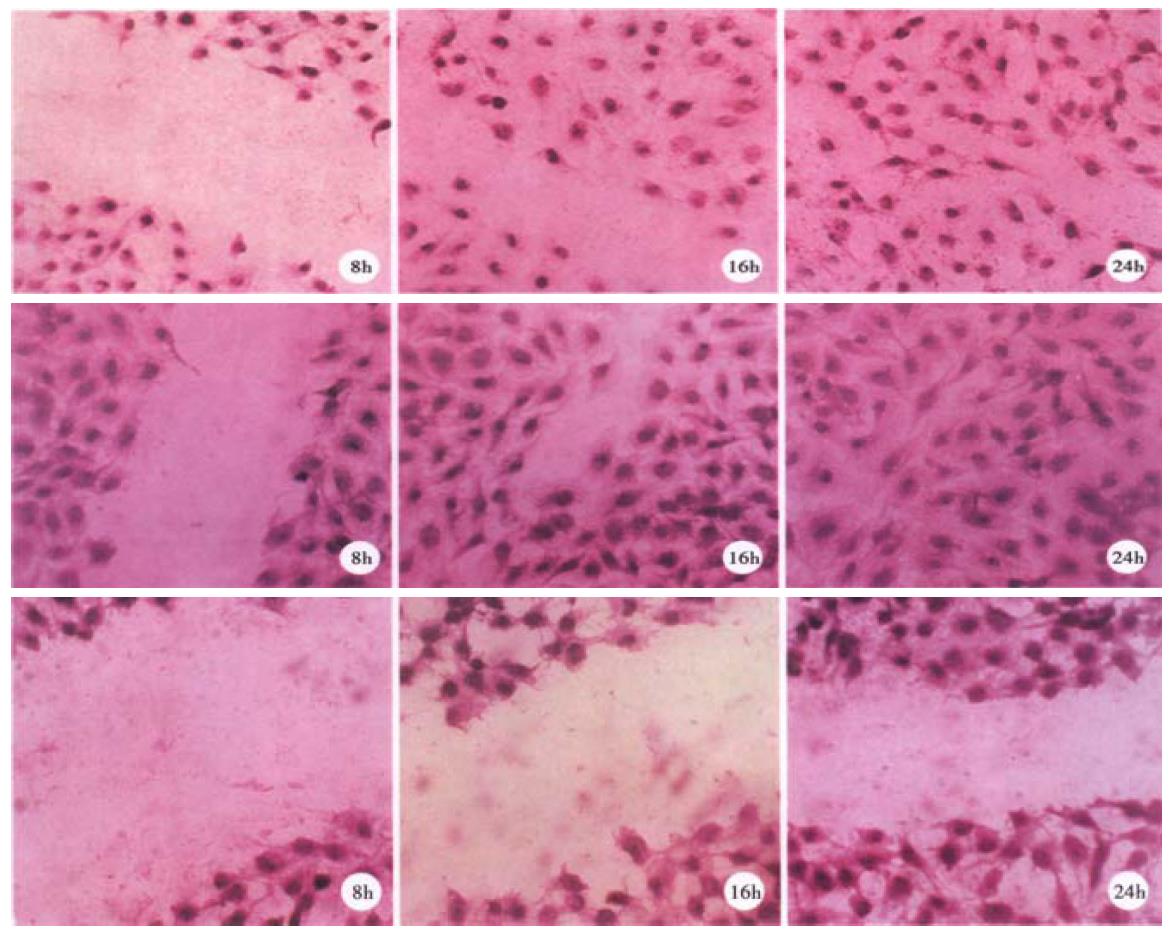
Figure 2 Migration ability of representative clones.
Subconfluent monolayers of the clones were "wounded" at time 0. The cells were allowed to migration into the cell-free area for 24 h then fixed and stained with crystal violet. A: RC1; B: RC2; C: SMMC 7721
- Citation: Wang Q, Lin ZY, Feng XL. Alterations in metastatic properties of hepatocellular carcinoma cell following H-ras oncogene transfection. World J Gastroenterol 2001; 7(3): 335-339
- URL: https://www.wjgnet.com/1007-9327/full/v7/i3/335.htm
- DOI: https://dx.doi.org/10.3748/wjg.v7.i3.335


