Copyright
©The Author(s) 2001.
World J Gastroenterol. Feb 15, 2001; 7(1): 9-15
Published online Feb 15, 2001. doi: 10.3748/wjg.v7.i1.9
Published online Feb 15, 2001. doi: 10.3748/wjg.v7.i1.9
Figure 1 Inhibitory effect of GA and acarbose on maltose absorption.
The intestinal loops in situ were perfused with 10 mmol/L maltose in the presence or absence of GA (1 g/L) and acarbose (mmol/L). The absorption of maltose is shown as percentage of maltose contained in the beginning of perfusion. Each point is expressed as mean ± SD of 5-10 determinations. (aP < 0.05, bP < 0.01)
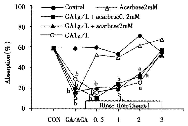
Figure 2 Alteration of maltose (10 mmol/L) absorption following application with GA and/or acarbose.
Each point shows the maltose absorption during 60 min perfusion of 10 mmol/L maltose following rise with Ringer’s solution after treatment of GA and/or acarbose (the second perfusion) except the points of “CON” or “GA/ACA” which shows the absorption during the first 1 hour perfusion with or without GA and acarbose (the first perfusion). The absorption of maltose is shown as percentage of maltose contained in the beginning of perfustion (aP < 0.05, bP < 0.01).
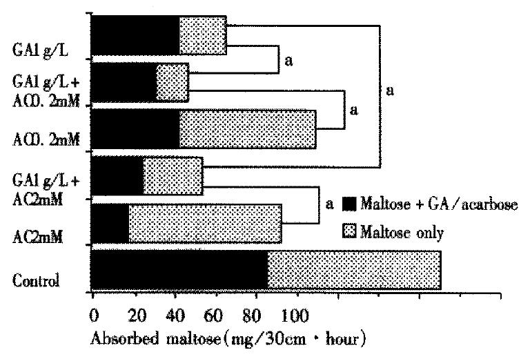
Figure 3 Maltose absorption during the two perfusions in the first 2.
5 hours. Maltose + GA/acarbose: first perfusion, 10 mmol/L maltose containing GA and/or acarbose was perfused for 1 hour. Maltose only: after first perfusion the intestinal loops were rinsed for 30 min, then 10 mmol/L maltose only was perfused for 1 hour again. The absorbed maltose is shown as the absorption in each perfusion. There was significant difference between each treated group versus control. (aP < 0.05, n = 5-10).
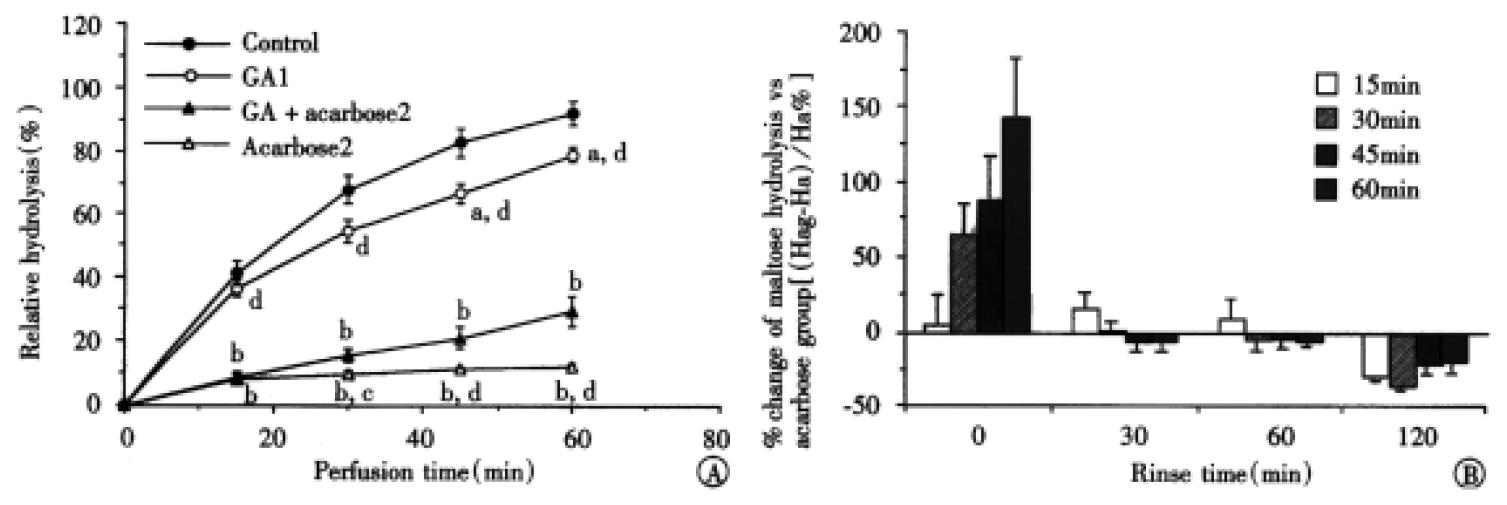
Figure 4 The dual effects of GA (1 mg/mL) on the activity of acarbose during the first perfusion (A), maltose with GA and/or acarbose was presented in the perfusates.
Maltose contained in the fluid at perfusion starting point was taken as 100%. aP < 0.05 and bP < 0.01 vs control; cP < 0.05 and dP < 0.01 vs GAa2. (n = 6-10) and second perfusion (B), the intestinal loops were rinsed for 30 min to 120 min, then 10 mmol/L maltose only was perfused for 1 h again. Each bar shows the percentage change of maltose hydrolysis (Hc%) in GAa2 group versus 2 mmol/L acarbose group in different perfusing times, which was calculated by the following equation: Hc% = (Hag-Ha)/Ha × 100%, where Hag and Ha represent, respectively, the hydrolyzed maltose in GAa2 group and in 2 mmol/L acarbose group. Hydrolysis of maltose in the acarbose group is believed as 100% (n = 3-10). A diminished effect in the beginning and improved effect in the end were observed.
Figure 5 The relationship of GA’s inhibitory effects on absorption of the maltose and on mobility of the intestinal ring.
A, an example of the inhibitory effect of GA (0.8 g/L) on the intestinal auto-rhythmic contraction. B, dose-dependently inhibitory effect of GA on amplitude of the small intestinal rings which is expressed as the tension induced by the auto-rhythmic contraction in the present different doses of GA. C, correlation of GA’s inhibitory effects on absorption of maltose and on tension of the intestinal ring. Each point comes from one dose of GA. The tension is calculated from the function of curve in the Figure 5B.
- Citation: Luo H, Wang LF, Imoto T, Hiji Y. Inhibitory effect and mechanism of acarbose combined with gymnemic acid on maltose absorption in rat intestine. World J Gastroenterol 2001; 7(1): 9-15
- URL: https://www.wjgnet.com/1007-9327/full/v7/i1/9.htm
- DOI: https://dx.doi.org/10.3748/wjg.v7.i1.9


