Copyright
©The Author(s) 2001.
World J Gastroenterol. Feb 15, 2001; 7(1): 60-65
Published online Feb 15, 2001. doi: 10.3748/wjg.v7.i1.60
Published online Feb 15, 2001. doi: 10.3748/wjg.v7.i1.60
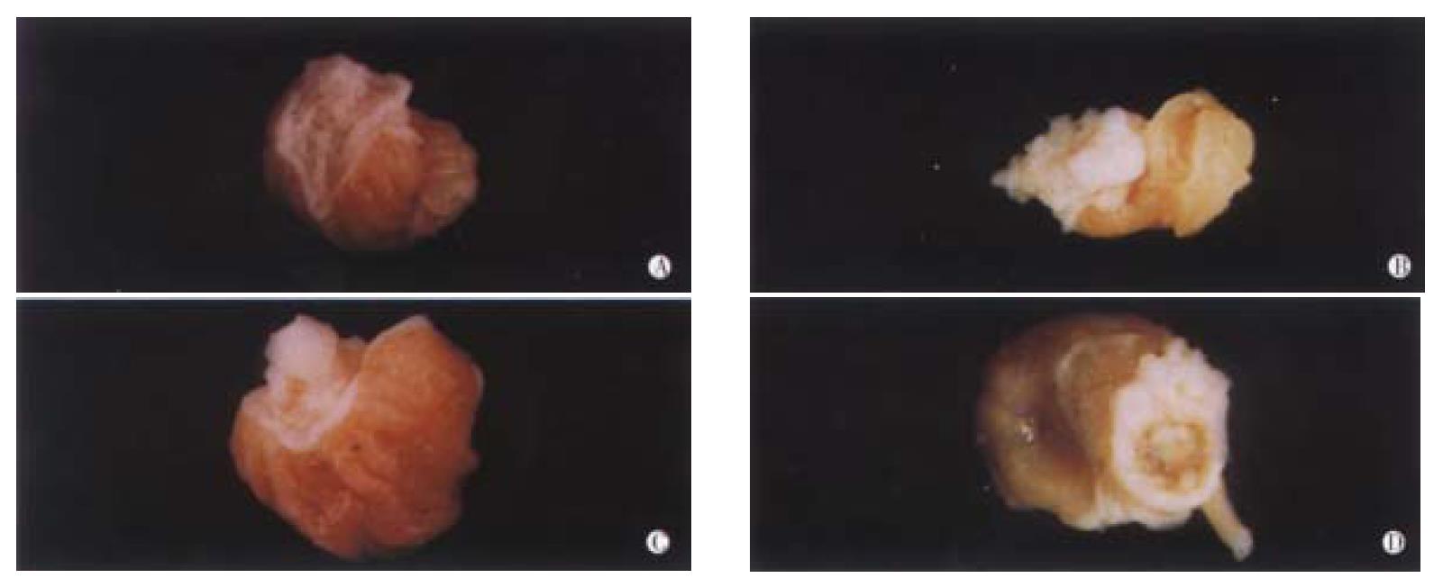
Figure 1 The model of B(a)P-induced forestomach tumor.
A. Normal mucosa of forestomach; B. Cauliflower-like tumor; C. Papilla-like tumor; D. Ulcer cavities-like tumor.
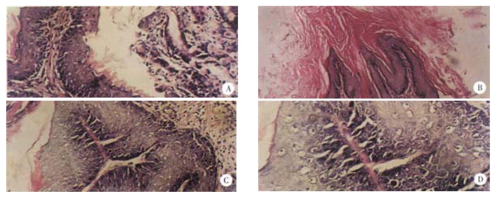
Figure 2 Pathological observation of B(a)P-induced forestomach tumor (HE staining) in mice.
A. The normal gastric mucosa × 200; B. The epithelium of gastric mucosa formed papillary projection × 100; C. Squamous tumor cells proliferated into connective tissues and they were polyhedral × 100; D. The mitotic figures (white arrow) in the squamous cell carcinoma × 400
- Citation: Wu K, Shan YJ, Zhao Y, Yu JW, Liu BH. Inhibitory effects of RRR-α-tocopheryl succinate on benzo(a)pyrene (B(a)P)-induced forestomach carcinogenesis in female mice. World J Gastroenterol 2001; 7(1): 60-65
- URL: https://www.wjgnet.com/1007-9327/full/v7/i1/60.htm
- DOI: https://dx.doi.org/10.3748/wjg.v7.i1.60


