Copyright
©The Author(s) 2001.
World J Gastroenterol. Feb 15, 2001; 7(1): 120-125
Published online Feb 15, 2001. doi: 10.3748/wjg.v7.i1.120
Published online Feb 15, 2001. doi: 10.3748/wjg.v7.i1.120
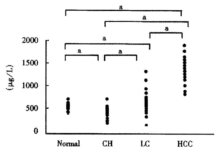
Figure 1 Serum concentration of sICAM-1 in normal controls (n = 50), in patients with chronic hepatitis (CH, n = 62), cirrhosis (LC, n = 60), and hepatocellular carcinoma (HCC, n = 151).
aP < 0.05, bP < 0.01.
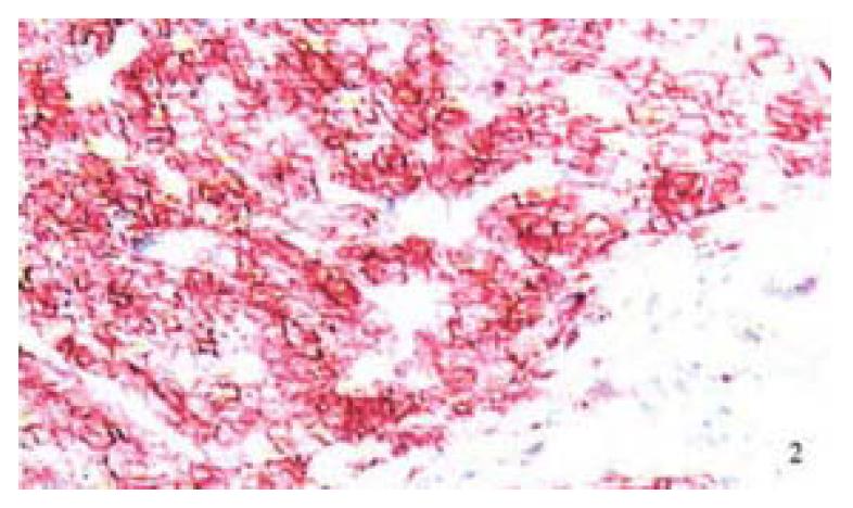
Figure 2 Immunohistochemical staining of ICAM-1 in hepatocellular carcinoma tissue is positive on the surface of tumor cells and in a honeycomb pattern.
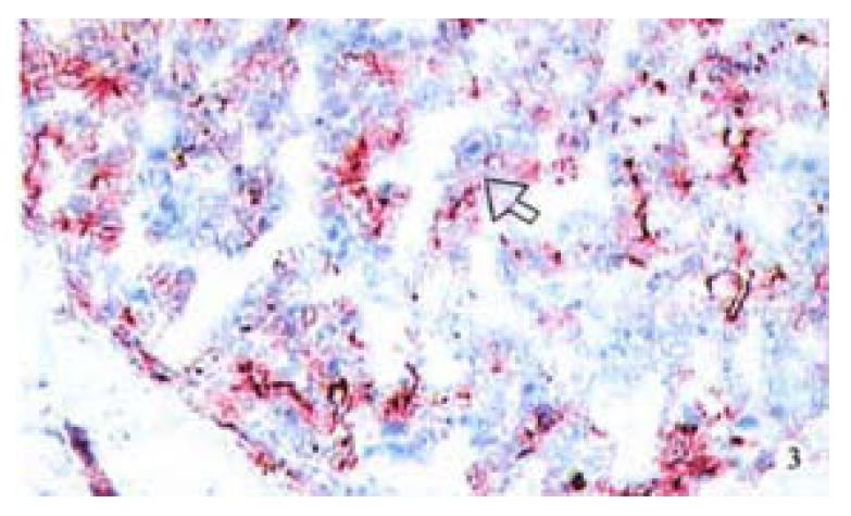
Figure 3 Increased expression of ICAM-1 in paracancerous tissue was shown with severe cirrhosis by immunohistochemical staining.
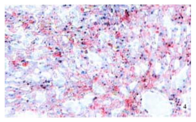
Figure 4 ICAM-1 expression was negative in normal liver tissue by immunohistochemical staining (AEC method).
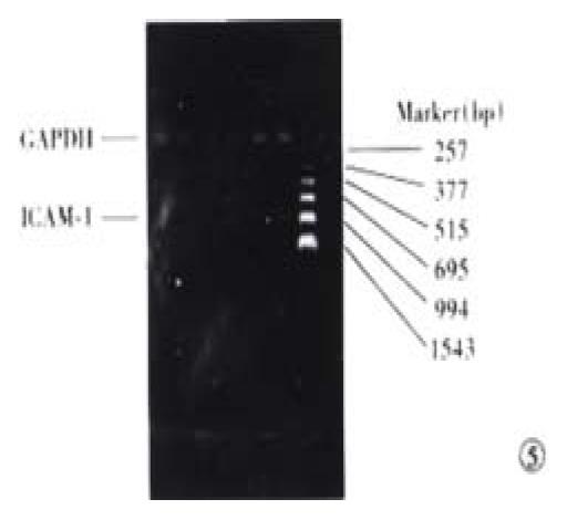
Figure 5 RT-PCR product gel electrophoresis.
Three ìL RT-PCR products of ICAM-1 and GAPDH run on 1.5% agarose gel stained with EB. Lane 1 and Lane 4: the control for ICAM-1 and GAPDH. Lane 2 and 5: paracarcinomatous tissue for ICAM-1 and GAPDH. Lane and 6: HCC tissues for ICAM-1 and GAPDH.
- Citation: Xu J, Mei MH, Zeng SE, Shi QF, Liu YM, Qin LL. Expressions of ICAM-1 and its mRNA in sera and tissues of patients with hepatocellular carcinoma. World J Gastroenterol 2001; 7(1): 120-125
- URL: https://www.wjgnet.com/1007-9327/full/v7/i1/120.htm
- DOI: https://dx.doi.org/10.3748/wjg.v7.i1.120


