Copyright
©The Author(s) 2000.
World J Gastroenterol. Dec 15, 2000; 6(6): 902-905
Published online Dec 15, 2000. doi: 10.3748/wjg.v6.i6.902
Published online Dec 15, 2000. doi: 10.3748/wjg.v6.i6.902
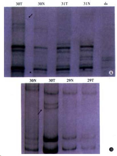
Figure 1 Patient of No.
30 positive for MSI. A: Lane N: corresponding normal tissue, Lane T: tumor tissue, Lane ds: di-strand control. B: MSI is defined as showing the presence of extra or shifting bands in PCR products using tumor DNA that are not visible in the products from corresponding normal tissue. (Left) D17D250, (Right) D2S123.
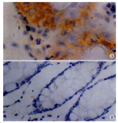
Figure 2 Immunohistochemistry of hMLH1.
(Left) Patient positive for hMLH1 immunostaining. × 40. (Right) Patient negative for hMLH1 immunostaining, × 20.
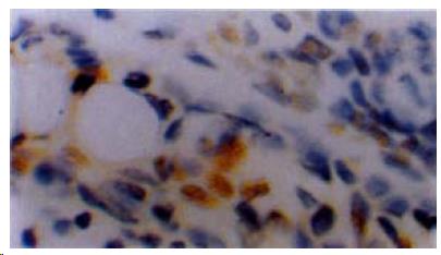
Figure 3 Immunostaining for PCNA in colorectal cancer tissue of patient with familial predisposition.
× 40.
- Citation: Wu BP, Zhang YL, Zhou DY, Gao CF, Lai ZS. Microsatellite instability, MMR gene expression and proliferation kinetics in colorectal cancer with famillial predisposition. World J Gastroenterol 2000; 6(6): 902-905
- URL: https://www.wjgnet.com/1007-9327/full/v6/i6/902.htm
- DOI: https://dx.doi.org/10.3748/wjg.v6.i6.902


