Copyright
©The Author(s) 2000.
World J Gastroenterol. Dec 15, 2000; 6(6): 898-901
Published online Dec 15, 2000. doi: 10.3748/wjg.v6.i6.898
Published online Dec 15, 2000. doi: 10.3748/wjg.v6.i6.898
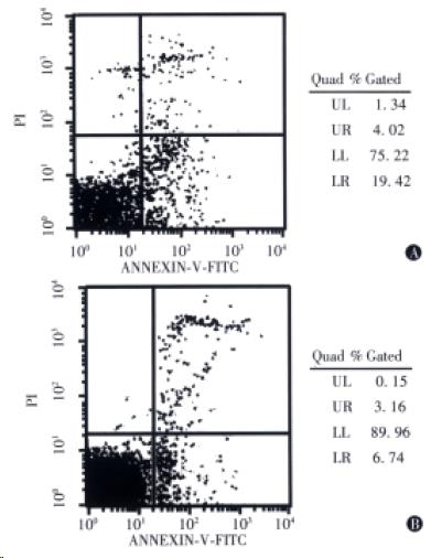
Figure 1 Bivariate AV/PI analysis of the gastric carcinoma (A) and adjacent non-neoplastic (B) tissues.
The different labeling patterns in this assay identify the different cell subpopulations. i.e. region LL, viable cells (AV-/PI-), region LR, apoptotic cells (AV+/PI-), region UR, dead cells (AV+/PI+).
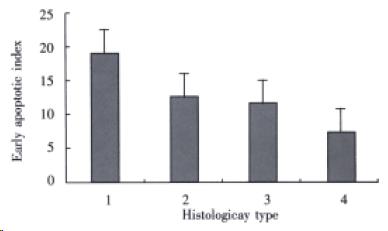
Figure 2 Early apoptosis in diffuse and intestinal gastric carcinomas.
1 and 2 were EAIs in carcinomatous tissues of diffuse and intestinal tumor, 19.0% ± 3.9% and 12.0% ± 4.3% (P = 0.0002); 3 and 4 were in adjacent non-neoplastic tissues, 10.8% ± 3.3% and 7.3% ± 4.2% (P = 0.0516).
- Citation: Zhou HP, Wang X, Zhang NZ. Early apoptosis in intestinal and diffuse gastric carcinomas. World J Gastroenterol 2000; 6(6): 898-901
- URL: https://www.wjgnet.com/1007-9327/full/v6/i6/898.htm
- DOI: https://dx.doi.org/10.3748/wjg.v6.i6.898


