Copyright
©The Author(s) 2000.
World J Gastroenterol. Oct 15, 2000; 6(5): 770-772
Published online Oct 15, 2000. doi: 10.3748/wjg.v6.i5.770
Published online Oct 15, 2000. doi: 10.3748/wjg.v6.i5.770
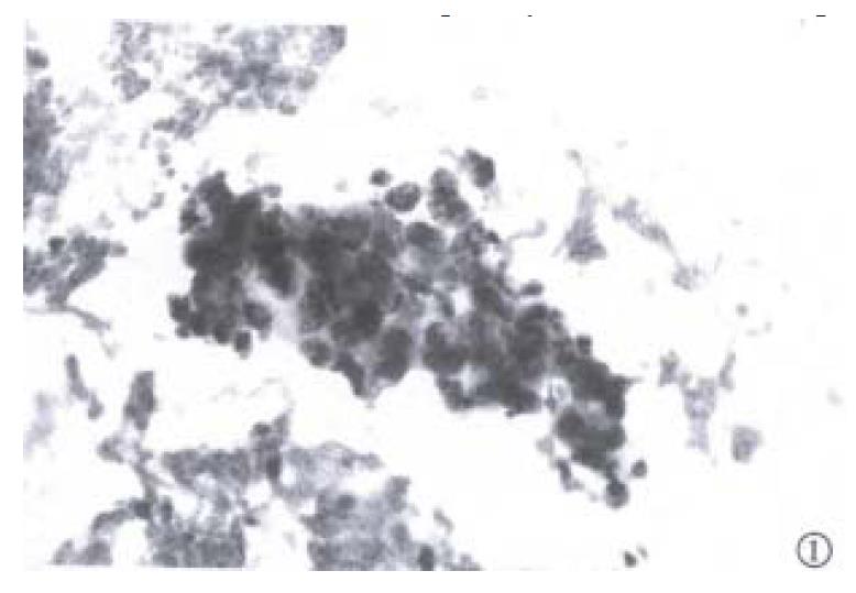
Figure 1 High power magnification of a cluster of cells from the axillart node aspirate, demonstrating pleomorphism and crowding o large poorly differentiated malignant cells.
Papanicolaou stain × 200
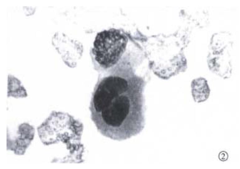
Figure 2 Single-lying cell from the axillary node aspirate, demonstrating high nuclear cytoplasmic ratios, central nuclei, and prominent nucleoli.
Diff-Quik × 400
- Citation: Alison MR, Leiman G, Kew MC. Metastasis in an axillary lymph node in hepatocellular carcinoma: a case report. World J Gastroenterol 2000; 6(5): 770-772
- URL: https://www.wjgnet.com/1007-9327/full/v6/i5/770.htm
- DOI: https://dx.doi.org/10.3748/wjg.v6.i5.770


