Copyright
©The Author(s) 2000.
World J Gastroenterol. Oct 15, 2000; 6(5): 651-658
Published online Oct 15, 2000. doi: 10.3748/wjg.v6.i5.651
Published online Oct 15, 2000. doi: 10.3748/wjg.v6.i5.651
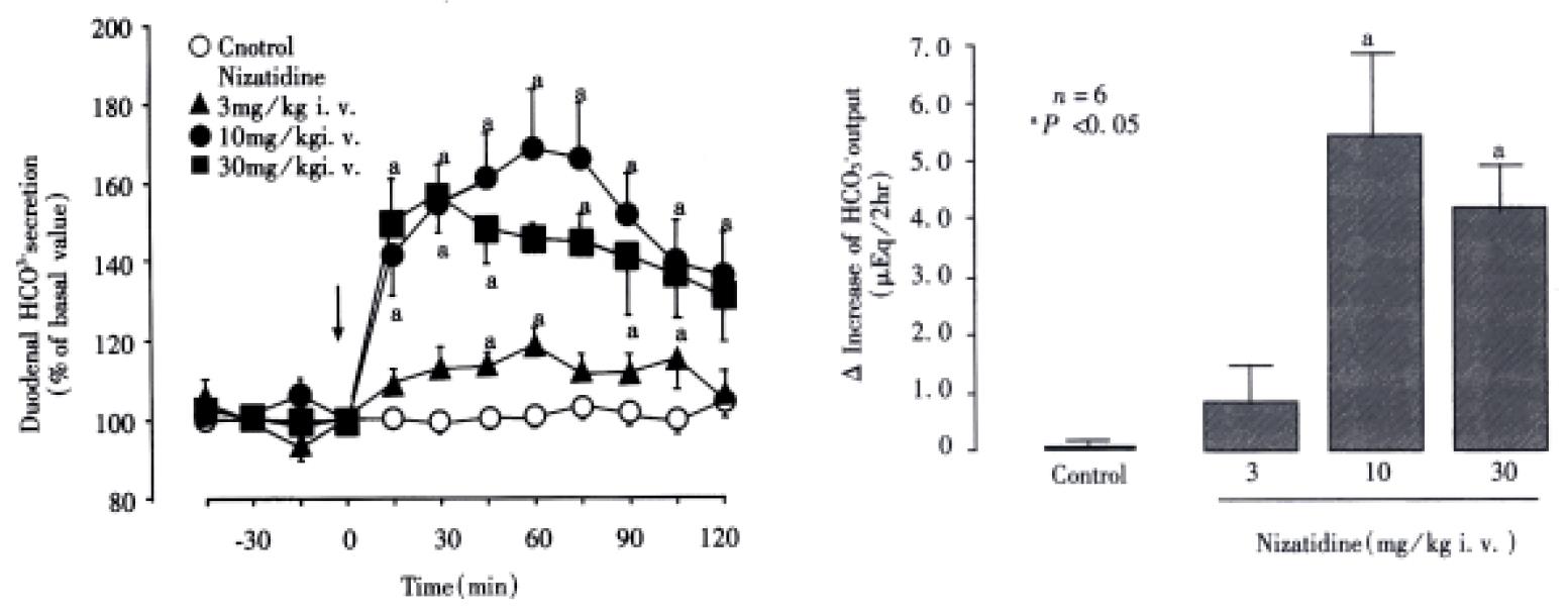
Figure 1 Effect of nizatidine on duodenal HCO3- secretion in rats.
The HCO3- secretion was measured by perfusing a duodenal loop with saline and by adding 10 mM HCl to the reservoir. Nizatidine (3-30 mg/kg) was administered i.v. as a single injection after the basal secretion had well stabilized, and the HCO3- secretion was measured f or 2 h. Data are expressed as % of basal values and represent the means ± SE of values determined every 15 min from 6 rats. Lower panel shows △increase of HCO3- output for 2 h. Data are presented as the means ± SE from 6 rats. aSignificant difference from controls, P < 0.05.
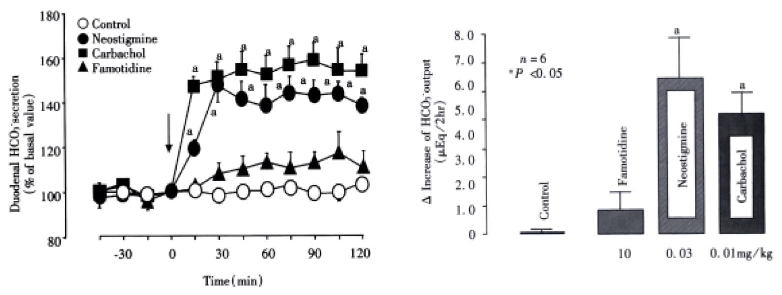
Figure 2 Effects of neostigmine, carbachol and famotidine on duodenal HCO3- secretion in rats.
The HCO3- secretion was measu red by perfusing a duodenal loop with saline and by adding 10 mM HCl to the reservoir. Neostigmine (0.03 mg/kg), carbachol (0.01 mg/kg) or famotidine (10 mg/kg) was administered i.v. as a single injection after the basal secretion had well stabilized, and the HCO3- secretion was measured for 2 h. Data are expressed as % of basal values and represent the means ± SE of values determined every 15 min from 6 rats. Lower panel shows △ increase of HCO3- output for 2 h. Data are presented as the means ± SE from 6 rats. aSignificant difference from controls, P < 0.05.
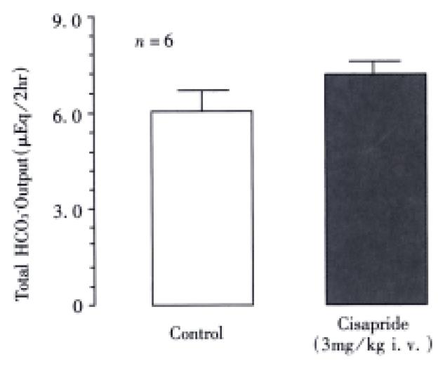
Figure 3 Effect of cisapride on duodenal HCO3- secretion in rats.
The HCO3- secretion was measured by perfusing a duodenal loop with saline and by adding 10 mM HCl to the reservoir. Cisapride (3 mg/kg) was administered i.v. as a single injection after the basal secretion had well stabilized, and the HCO3- secretion was measured for 2 h. Data indicate total HCO3- output obtained for 2 h after administration of cisapride, and represent the means ± SE from 6 rats.
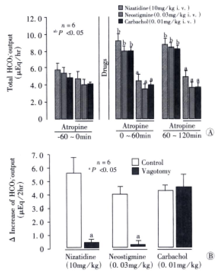
Figure 4 Effects of atropine or bilateral vagotomy on the HCO3- stimulatory action of nizatidine, neostigmine and carbachol in the rat duodenum.
The HCO3- secretion was measured by perfusing a duodenal loop with saline and by adding 10 mM HCl to the reservoir. Neostigmine (0.03 mg/kg), carbachol (0.01 mg/kg) or famotidine (10 mg/kg) was administered i.v. as a single injection after the basal secretion had well stabilized, and the HCO3- secretion was measured for 2 h. Atropine (1 mg/kg) was given s.c. 1 h before admin istration of the above agents. Bilateral vagotomy was performed acutely at the cervical portion 2 h before the administration of the agents. Data are presented as the means ± SE from 6 rats. Significant difference at P < 0.05; afrom corresponding groups without atropine; bfrom corresponding values observed for 1 h before the treatment (time: -60-0 min)
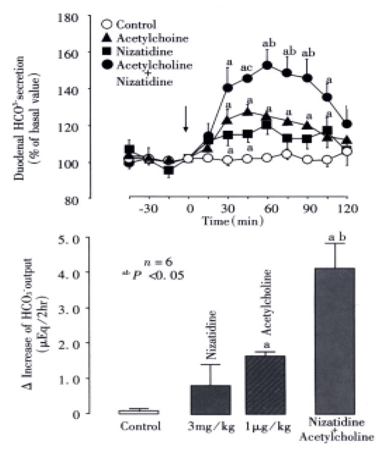
Figure 5 Effects of nizatidine and acetylcholine, either alone or in combination, on duodenal HCO3- secretion in rats.
The HCO3- secretion was measured by perfusing a duodenal loop with saline and by adding 10 mM HCl to the reservoir. Nizatidine (3 mg/kg) and acetylcholine (0.003 mg/kg), either alone or in combination, were administered i.v. after the basal secretion had well stabilized, and the HCO3- secretion was measured for 2 h. Data are expressed as % of basal values and represe nt the means ± SE of values determined every 15 min from 6 rats. Lower panel shows △increase of HCO3- output for 2 h. Data are presented as the means ± SE from 6 rats. Significant difference at P < 0.05a from control; bfrom nizatidine or acetylcholine.
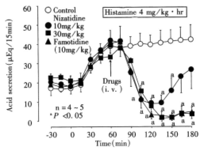
Figure 6 Effects of nizatidine and famotidine on histamine-induced gastric acid secretion in rats.
A rat stomach mounted in an ex-vivo chamber was perfused with saline, and the acid secretion was measur ed by adding 100 mM NaOH to the reservoir. The acid secretion was stimulate d by i.v. infusion of histamine (4 mg/kg/h), while nizatidine (10 and 30 mg/kg) or famotidine (10 mg/kg) was administered i.v. as a single injection 1 h after the onset of histamine infusion, when the acid secretion had reached a plateau. Data are presented as the means ± SE of values determined every 15 min from 5 rats. aSignificant difference from controls, P < 0.05.
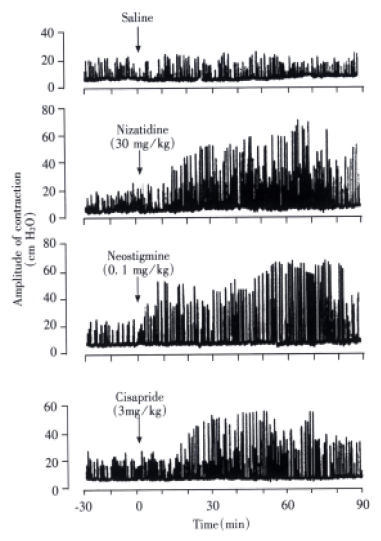
Figure 7 Representative recordings of gastric motor activity in conscious rats, before and after administration of nizatidine.
gastric motor activity was monitored as intraluminal pressure recordings, by using a miniature balloon placed in the stomach. Nizatidine (30 mg/kg) was administered i.v. as a single injection after the basal motility had well stabilized. Atropine (1 mg/kg) was given s.c. 1 h after administration of nizatidine. Note that gastric motor activity was apparently enhanced by nizatidine at the doses that stimulate HCO3- secretion in the rat duodenum, and this action was completely blocked by atropine.
- Citation: Takeuchi K, Kawauchi S, Araki H, Ueki S, Furukawa O. Stimulation by nizatidine, a histamine H2-receptor antagonist, of duodenal HCO3- secretion in rats: relation to anti-cholinesterase activity. World J Gastroenterol 2000; 6(5): 651-658
- URL: https://www.wjgnet.com/1007-9327/full/v6/i5/651.htm
- DOI: https://dx.doi.org/10.3748/wjg.v6.i5.651


