Copyright
©The Author(s) 2000.
World J Gastroenterol. Aug 15, 2000; 6(4): 608-612
Published online Aug 15, 2000. doi: 10.3748/wjg.v6.i4.608
Published online Aug 15, 2000. doi: 10.3748/wjg.v6.i4.608
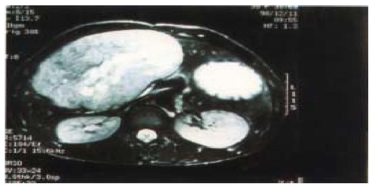
Figure 1 MRI image of case 7.
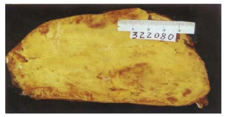
Figure 2 Macroscopic appearance of AML with yellowish fatty areas (case 7).
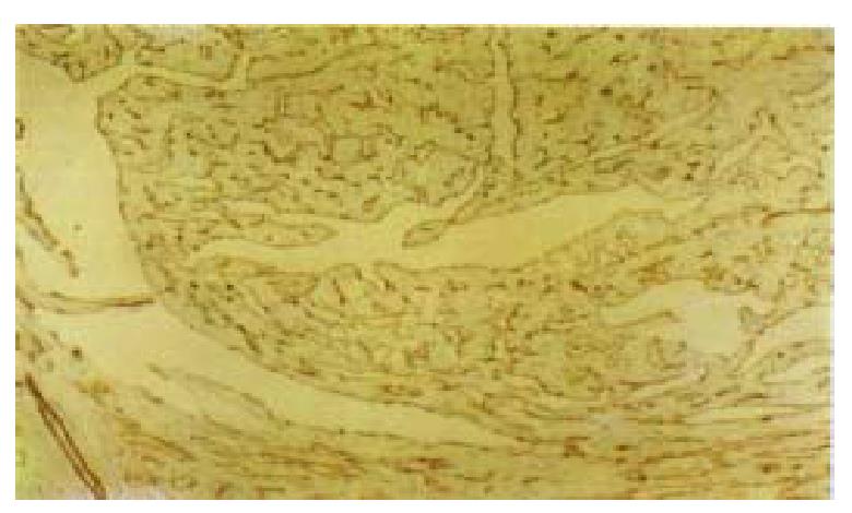
Figure 3 Thin-walled vessles and trabecular tumor cells of AML.
IH: CD31 × 100
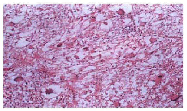
Figure 4 Multinucleus cells in a large number in AML of case 9.
HE × 100
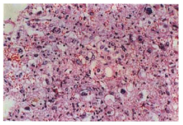
Figure 5 Pleomorphistic and large bizarre cells.
HE × 200
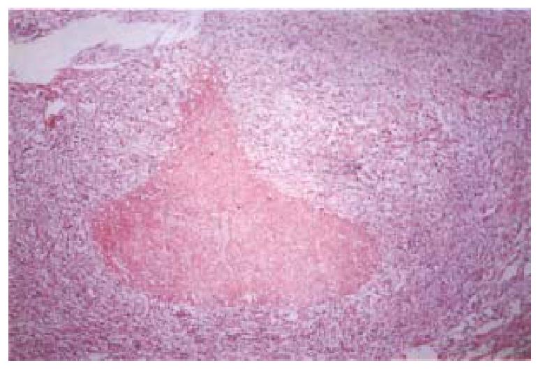
Figure 6 Local necrotic area of case 9.
HE × 50
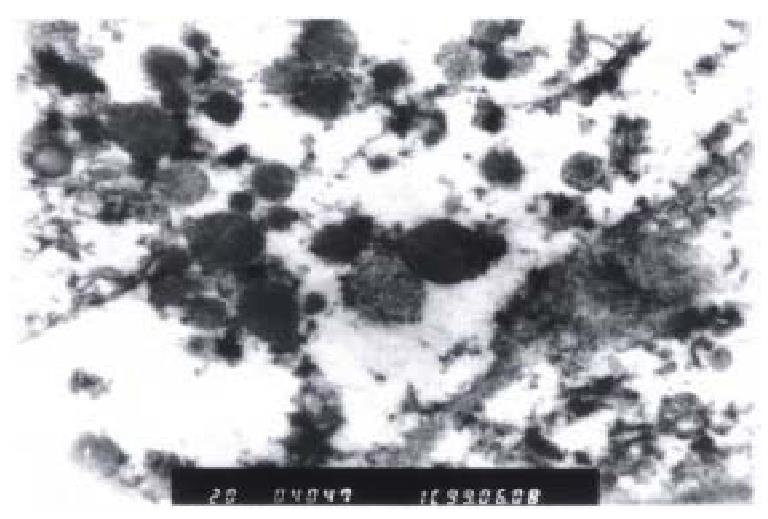
Figure 7 Ultrastructure of neoplastic tissue: glycogen, electron-dense granules.
EM × 20000
- Citation: Zhong DR, Ji XL. Hepatic angiomyolipoma-misdiagnosis as hepatocellular carcinoma: A report of 14 cases. World J Gastroenterol 2000; 6(4): 608-612
- URL: https://www.wjgnet.com/1007-9327/full/v6/i4/608.htm
- DOI: https://dx.doi.org/10.3748/wjg.v6.i4.608


