Copyright
©The Author(s) 2000.
World J Gastroenterol. Aug 15, 2000; 6(4): 546-552
Published online Aug 15, 2000. doi: 10.3748/wjg.v6.i4.546
Published online Aug 15, 2000. doi: 10.3748/wjg.v6.i4.546
Math 1 Math(A1).
Math 2 Math(A1).
Figure 1 Expression of CD45 and ANK61 antigen on hepatic NK cells (pit cells).
The cells were washed out of the liver by sinusoidal lavage. After Ficoll-Paque gradient centrifugation and nylon-wool adherence to remove erythrocytes, granul ocytes, monocytes and B lymphocytes, the cells were stained with anti-NKR-P1A mAb (3.2.3) (phycoerythrin) and anti-ANK61 antigen (ANK61), anti-CD45 mAb (ANK 74) (fluorescein) and analyzed by two-color flow cytometry. CD45 and ANK61 anti gen were expressed on the x-axis (fluorescein), NKR-P1A on the y-axis (phycoe rythrin).
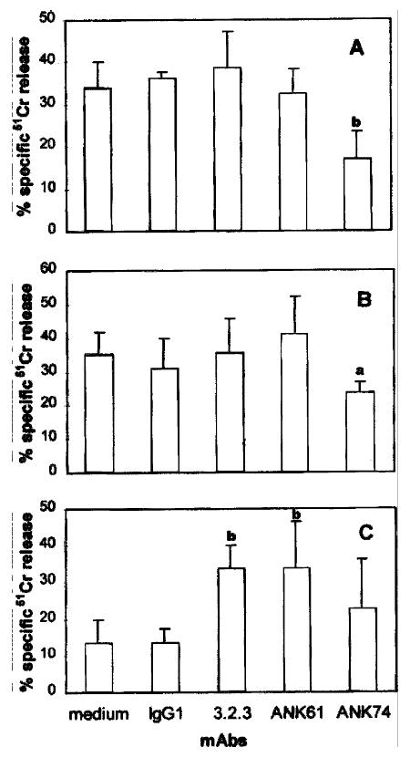
Figure 2 Effect of the mAbs on pit cell-mediated CC531s (A), YAC-1 (B) and P81 5 (C) target cell cytolysis.
51Cr-labeled target cells were incubated at an E:T ratio of 10:1 with freshly isolated pit cells in the absence or prese nce of the mAbs. (A), Cytolysis of CC531s cells was measured in an 18h 51Cr-release assay. (B), Cytolysis of YAC-1 cells was measured in a 4-h 51Cr-release assay. (C), Cytolysis of P815 cells was measured in a 4-h 51Cr-release assay. Values are mean ± SD from four different experiments. aP < 0.05, bP < 0.01 vs the control
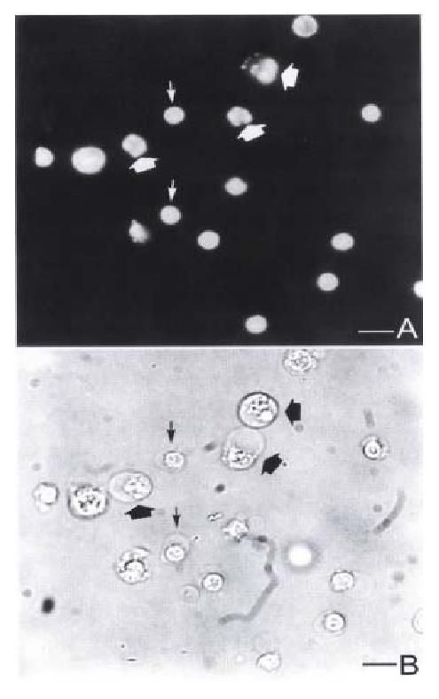
Figure 3 Fluorescence and light micrographs of YAC-1 cells coincubated with pit cells at an E:T ratio of 10:1 for 3 h.
(A) Fluorescence micrograph showing the apoptotic YAC-1 cells with fragmented nuclei (thick arrows) and pit cells ( thin arrows). Light micrograph shows the same field as (A). Bar = 5 μm.
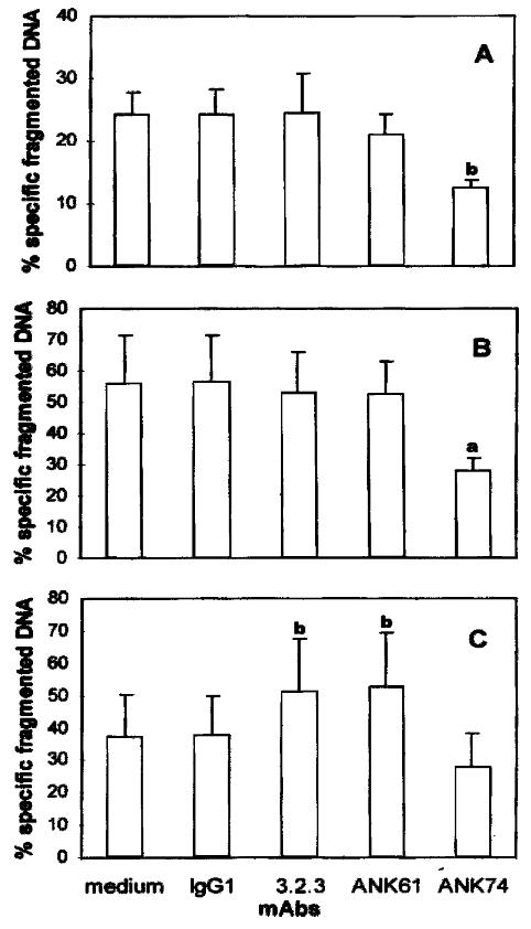
Figure 4 Effect of the mAbs on pit cell-induced CC531s (A), YAC-1 (B) and P8 15 (C) target cell apoptosis.
[3H]-TdR labeled target cells were incubated at an E:T ratio of 10:1 with freshly isolated pit cells for 3 h in the absence or presence of the mAbs. Values are mean ± SD from four different ex periments.aP < 0.05, bP < 0.01 vs the control.
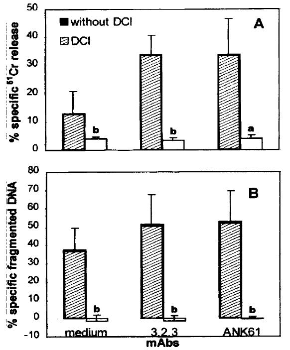
Figure 5 Effect of DCI on mAbs 3.
2.3 and ANK61-enhanced Fcγ R+ P815 cell cytolysis (A) and apoptosis (B) by pit cells. Pit cells were preincubated with 50 μmol/L DCI for 30 min at 37 °C. After washing twice, the cells were coincubated with P815 cells as mentioned in materials and methods. Values are mean ± SD from three different experiments. aP < 0.05, bP < 0.01 vs the corresponding control.
- Citation: Luo DZ, Vermijlen D, Ahishali B, Triantis V, Vanderkerken K, Kuppen PJ, Wisse E. Participation of CD45, NKR-P1A and ANK61 antigen in rat hepatic NK cell (pit cell)-mediated target cell cytotoxicity. World J Gastroenterol 2000; 6(4): 546-552
- URL: https://www.wjgnet.com/1007-9327/full/v6/i4/546.htm
- DOI: https://dx.doi.org/10.3748/wjg.v6.i4.546


