Copyright
©The Author(s) 2000.
World J Gastroenterol. Aug 15, 2000; 6(4): 522-525
Published online Aug 15, 2000. doi: 10.3748/wjg.v6.i4.522
Published online Aug 15, 2000. doi: 10.3748/wjg.v6.i4.522
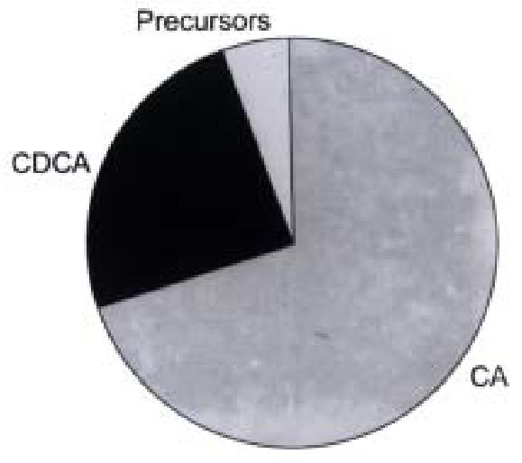
Figure 1 Distribution of cholic acid, chenodeoxycholic acid and precursors formed in primary cultures of human hepatocytes.
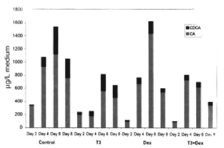
Figure 2 Formation of cholic acid and chenodeoxycholic acid in primary cultures of human hepatocytes during d 2, 4, 6 and 8.
Effects of adding T3 and dexa methasone (DEX) alone and in combination.
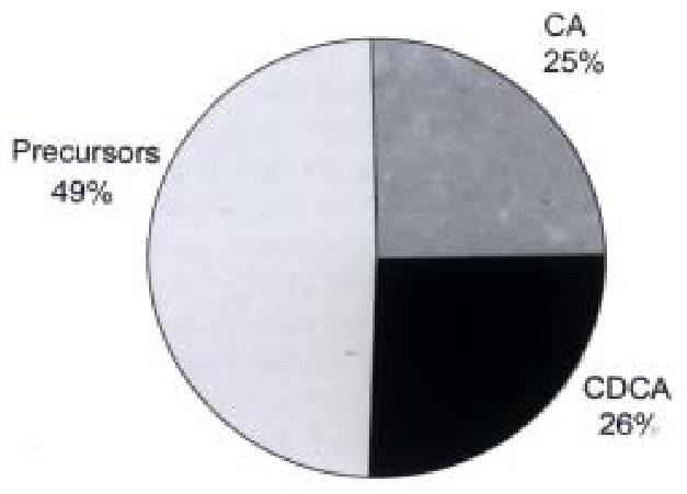
Figure 3 Distribution of cholic acid, chenodeoxycholic acid and precursors formed in cultures of HepG2 cells (data from ref.
7).
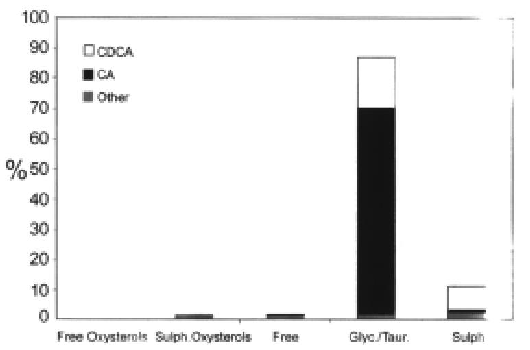
Figure 4 Conjugation of bile acids and precursors formed in primary cultures of human hepatocytes.
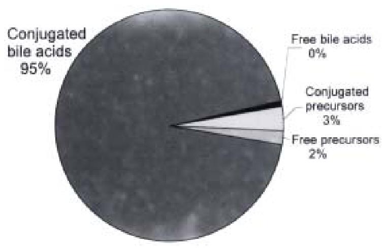
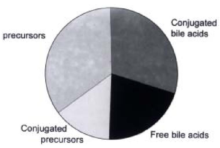
Figure 5 Distribution of free and conjugated bile acids and potential interme diates isolated from medium on the fifth day of incubating primary human hepatoc ytes.
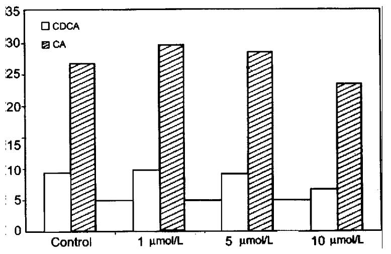
CDCA = chenodeoxycholic acid; CA = cholic acid.
Figure 6 Conjugation of bile acids and precursors formed in cultures of HepG2 cells (data from ref.
7).
Figure 7 Effect of cyclosporin A on bile acid formation in primary human hepa tocytes.
- Citation: Einarsson C, Ellis E, Abrahamsson A, Ericzon BG, Björkhem I, Axelson M. Bile acid formation in primary human hepatocytes. World J Gastroenterol 2000; 6(4): 522-525
- URL: https://www.wjgnet.com/1007-9327/full/v6/i4/522.htm
- DOI: https://dx.doi.org/10.3748/wjg.v6.i4.522


