Copyright
©The Author(s) 2000.
World J Gastroenterol. Jun 15, 2000; 6(3): 461-464
Published online Jun 15, 2000. doi: 10.3748/wjg.v6.i3.461
Published online Jun 15, 2000. doi: 10.3748/wjg.v6.i3.461
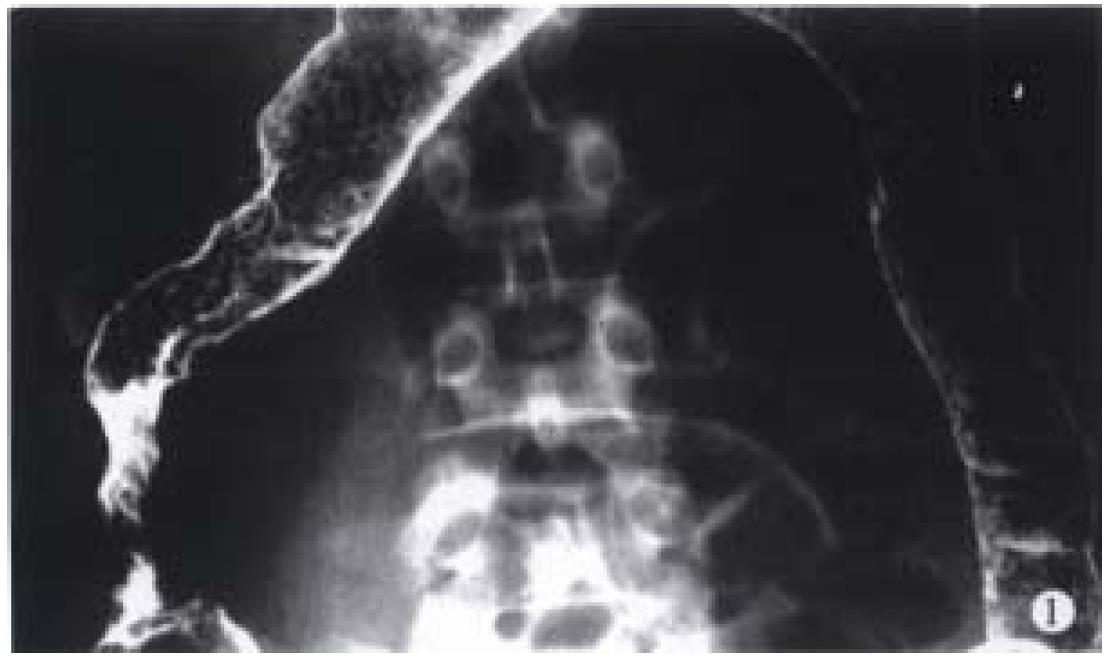
Figure 1 Barium enema examination perfoemed on March 18, 1993.
Stricture associated with cobblestone appearance was seen in the ascending colon. Small inflammatory polyps were observed in the transverse colon and descending colon.
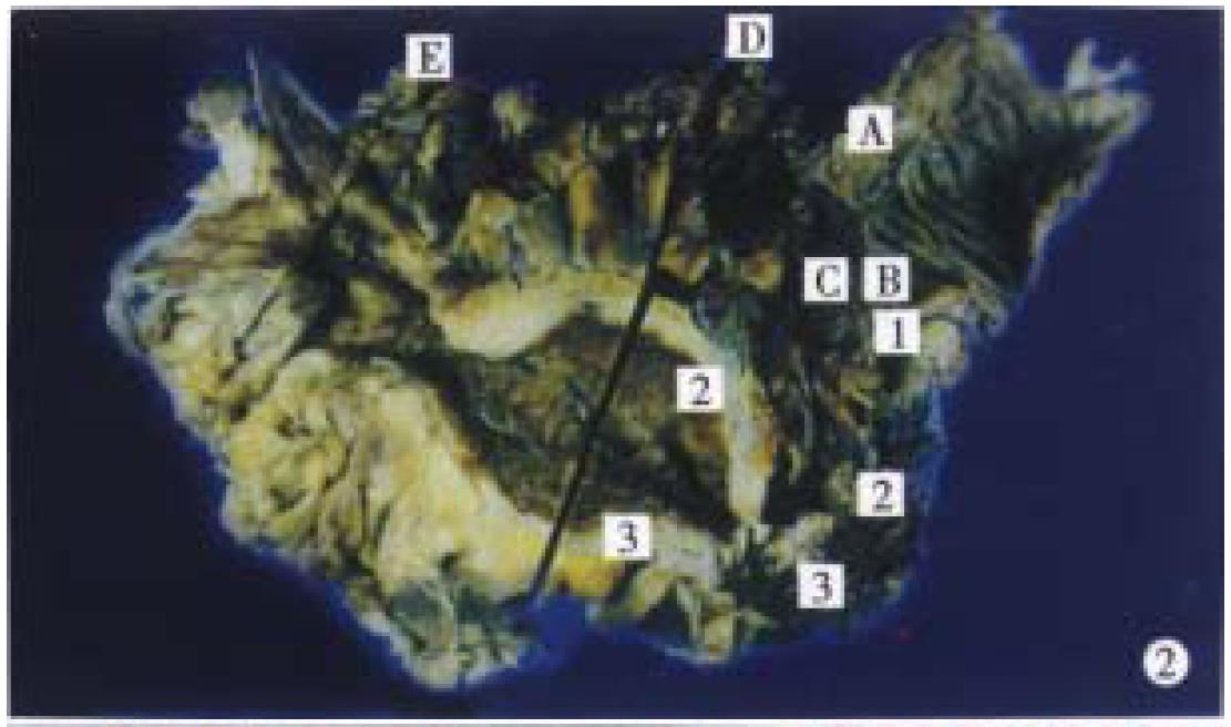
Figure 2 Surgical specimen of the terminal ileum, cecum, and ascending colon (May 26, 1993).
Thickening of the bowel wall, cobblestone appearance, and longitudinal ulceration were found, Histologically, transmur al inflammation and noncaseating epithelioid cell granuloma were found.
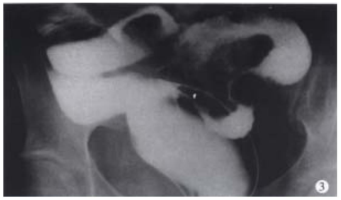
Figure 3 Gastrografin enema examination performed on May 8, 1997.
A stricture was seen around the ileosigmoid anastomosis.
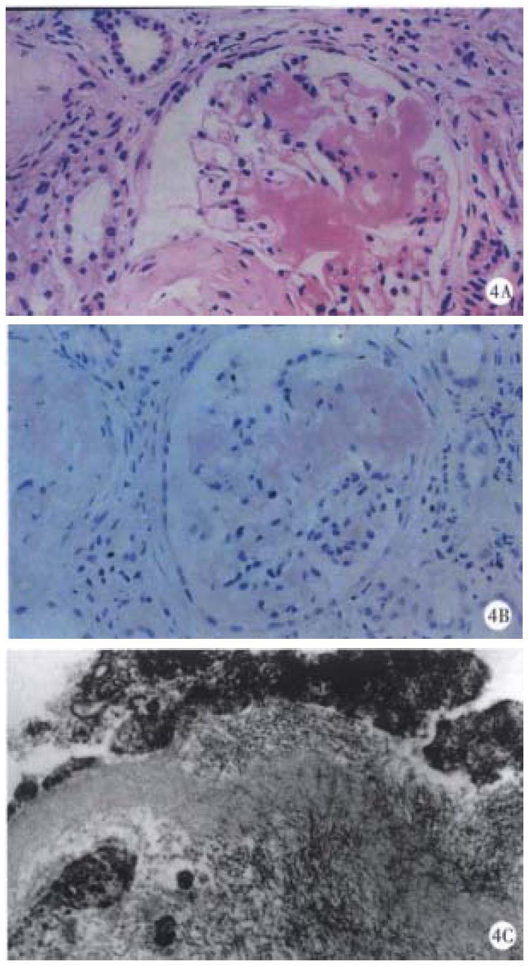
Figure 4 Findings of the renal biopsy.
A: Histological findings (hematocylin and eosin). Amorphous, eosin-stained deposits were seen in the mesangial areas. B: Histological findings (Congo red stain). The deposits were Congo red positive. Congo red stain showed reddish pink deposits that demonstrated apple-green birefringence when examined under polarized light. C: Electron microscopic findings. Fine fibrils (8 to 10 nm in diameter) arranged randomly or in bundles were found in the mesangium.
- Citation: Saitoh O, Kojima K, Teranishi T, Nakagawa K, Kayazawa M, Nanri M, Egashira Y, Hirata I, Katsu KI. Renal amyloidosis as a late complication of Crohn's disease: a case report and review of the literature from Japan. World J Gastroenterol 2000; 6(3): 461-464
- URL: https://www.wjgnet.com/1007-9327/full/v6/i3/461.htm
- DOI: https://dx.doi.org/10.3748/wjg.v6.i3.461


