Copyright
©The Author(s) 2000.
World J Gastroenterol. Apr 15, 2000; 6(2): 252-254
Published online Apr 15, 2000. doi: 10.3748/wjg.v6.i2.252
Published online Apr 15, 2000. doi: 10.3748/wjg.v6.i2.252
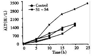
Figure 1 Serum ALT changes in FHF animals.
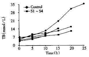
Figure 2 TB changes in FHF animals.
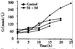
Figure 3 Serum Cr changes in FHF animals.
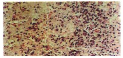
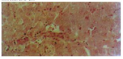
Figure 4 Liver biopsy specimens from control group.
Large area of necrosis. LM, × 200
Figure 5 Hepatic histology of support animals: mild liver necrosis.
LM, × 200
- Citation: Wang YJ, Li MD, Wang YM, Chen GZ, Lu GD, Tan ZX. Effect of extracorporeal bioartificial liver support system on fulminant hepatic failure rabbits. World J Gastroenterol 2000; 6(2): 252-254
- URL: https://www.wjgnet.com/1007-9327/full/v6/i2/252.htm
- DOI: https://dx.doi.org/10.3748/wjg.v6.i2.252


