Copyright
©The Author(s) 1999.
World J Gastroenterol. Oct 15, 1999; 5(5): 383-387
Published online Oct 15, 1999. doi: 10.3748/wjg.v5.i5.383
Published online Oct 15, 1999. doi: 10.3748/wjg.v5.i5.383
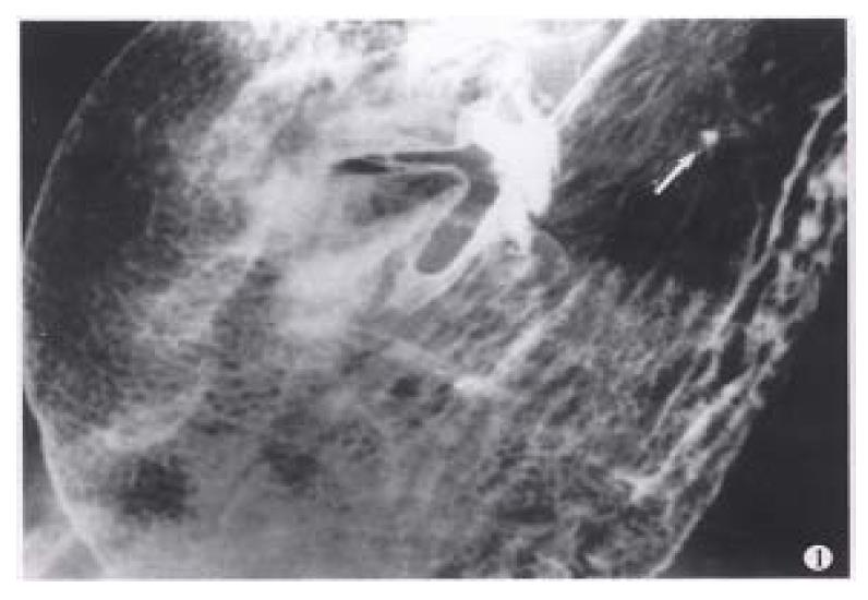
Figure 1 Areae gastricae.
Well-circumscribed polygonal radiolucencies surrounded by barium-filled shallow grooves are clearly seen on the mucosal surface. A small benign ulcer is also displayed (arrowhead).
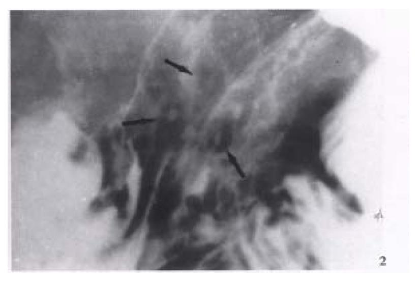
Figure 2 Erosive gastritis.
Multiple varioliform erosions (arrows) in the posterior wall of gastric body are seen as tiny barium collections with surrounding halos of edematous mucosa-“target” sign.
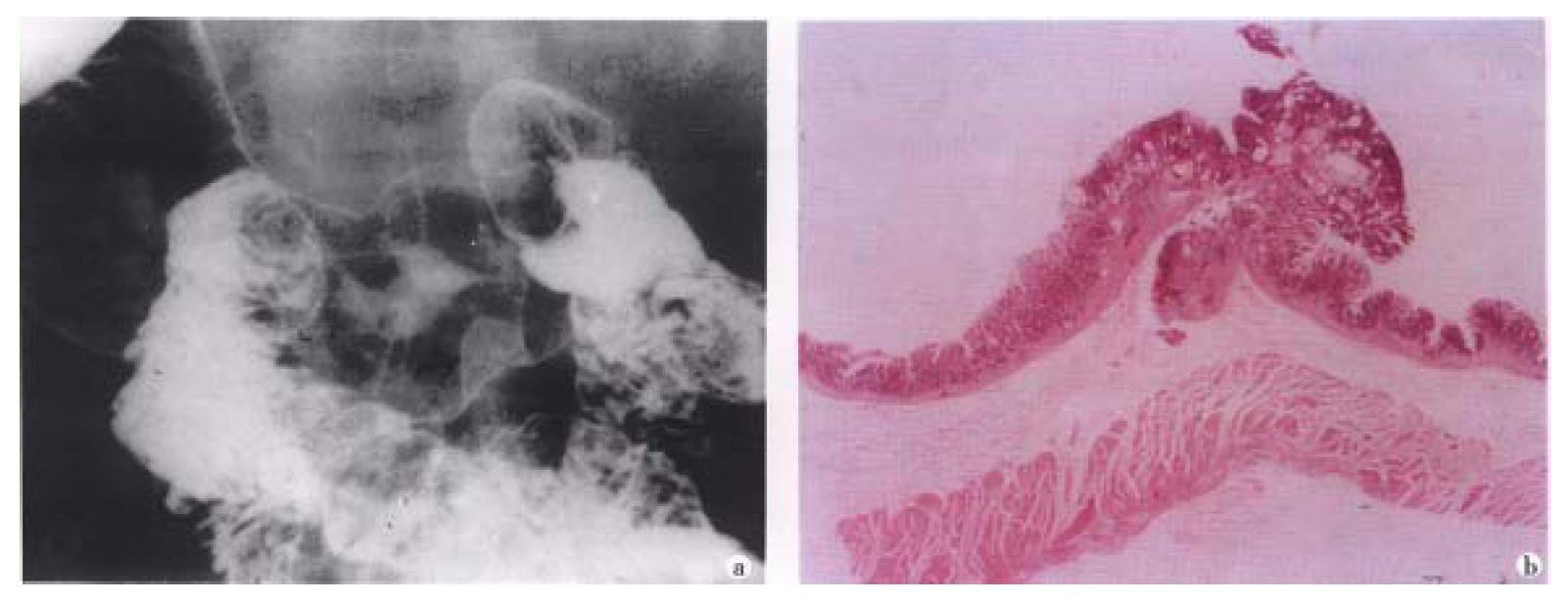
Figure 3 Small early gastric cancer (type I).
A sessile protruded lesion (5 mm in size) with its base puckered into the lumen is seen on the greater curvature of antrum.B. Micrograph shows the lesion originates from mucosa and infilt rates to the submucosal layer.
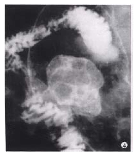
Figure 4 Gastric carcinoma (Borrmann’s type I).
A bulky cauliflower-like mass etched in white by a thin layer of barium is seen on the greater curvature of the stomach.
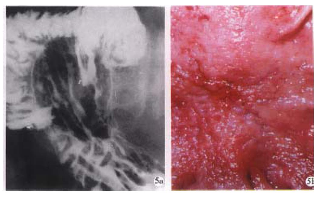
Figure 5 Linear ulcer.
A long linear ulcer paralleled to the lesser curvature (arrowhead) shown on the gastric posterior wall, representing the healing and healed stage of ulcer. B. Macrograph shows that the fine curve linear ulcer is composed of re-epithelialized (healed) and granulating part (healing).
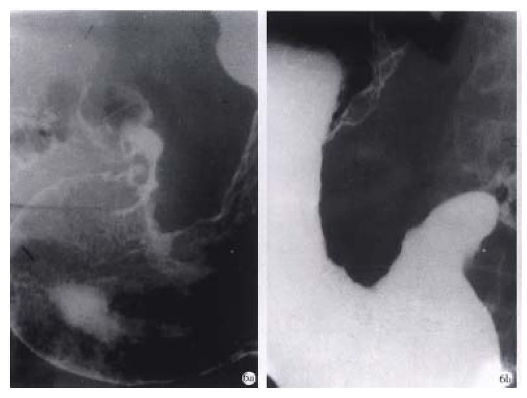
Figure 6 Advanced gastric carcinoma.
A. Double-contrast phase. B. Filling phase reveal marked “rigid” and “coarse” contour of the lesser curvature and lumen narrowing caused by infiltrative carcinoma.
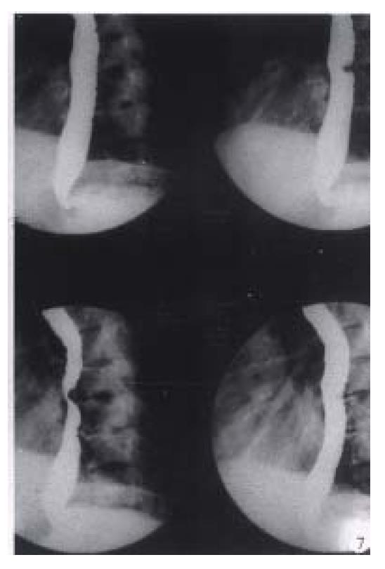
Figure 7 Digital esophageal barium radiography.
The defect on posterior aspect of middle esophagus caused by the proliferation of thoracic vertebrae is easily displayed by rapid sequence images (0.5 pictures/sec) and results in dysphagy clinically.
- Citation: Chen JR. Reassessment of barium radiographic examination in diagnosing gastrointestinal diseases. World J Gastroenterol 1999; 5(5): 383-387
- URL: https://www.wjgnet.com/1007-9327/full/v5/i5/383.htm
- DOI: https://dx.doi.org/10.3748/wjg.v5.i5.383


