Copyright
©The Author(s) 1998.
World J Gastroenterol. Oct 15, 1998; 4(5): 459-460
Published online Oct 15, 1998. doi: 10.3748/wjg.v4.i5.459
Published online Oct 15, 1998. doi: 10.3748/wjg.v4.i5.459
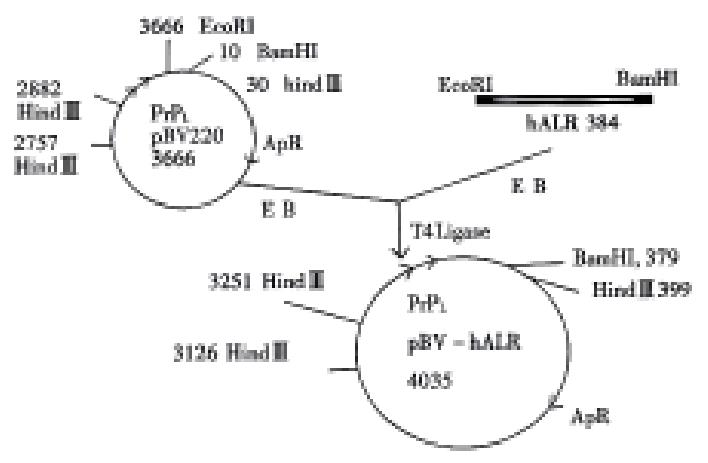
Figure 1 Construction pBV-hALR vector.
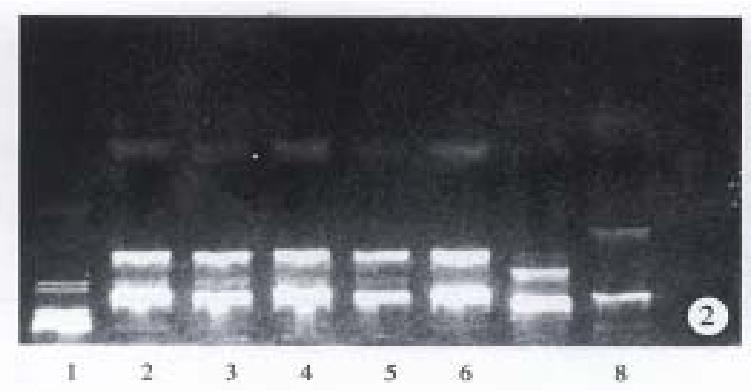
Figure 2 Detection pBV-hALR vector by Hind III.
1 γ/Hind III DNA marker 2-6 pBV-hALR vector digested by Hind III 8 pBV-220 vector digested by Hind III
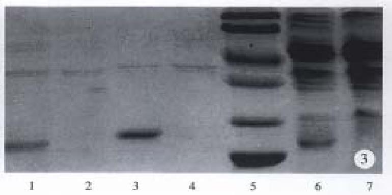
Figure 3 SDS-PAGE profile of expressed rhALR.
1 Precipitation of E.coli JM109 with pBV-hALR after sonication; 2 Precipitation of E.coli JM109 with pBV-220 after sonication; 3 Precipitation of E.coli DH5α with pBV-hALR after sonication; 4 Precipitation of E.coli DH5α with pET after sonication; 5 Protein standard (Mr 14 400, 21 500, 31 000, 45 000, 66 2000, 97000);6 E. coli JM109 with pBV-hALR 7 E. coli JM109 with pBV-220.
- Citation: Yi XR, Kong XP, Zhang YJ, Tong MH, Yang LP, Li RB. High expression of human augmenter of liver regeneration in E. coli. World J Gastroenterol 1998; 4(5): 459-460
- URL: https://www.wjgnet.com/1007-9327/full/v4/i5/459.htm
- DOI: https://dx.doi.org/10.3748/wjg.v4.i5.459


