Copyright
©The Author(s) 1998.
World J Gastroenterol. Apr 15, 1998; 4(2): 133-136
Published online Apr 15, 1998. doi: 10.3748/wjg.v4.i2.133
Published online Apr 15, 1998. doi: 10.3748/wjg.v4.i2.133
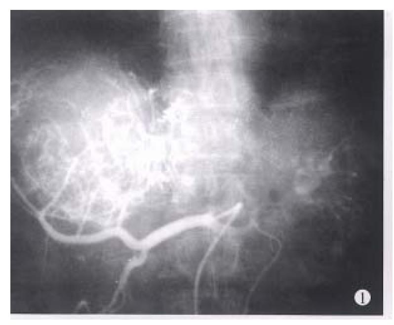
Figure 1 The hepatic arteriography shows a large tumor with hypervasculars in the right lobe of liver.
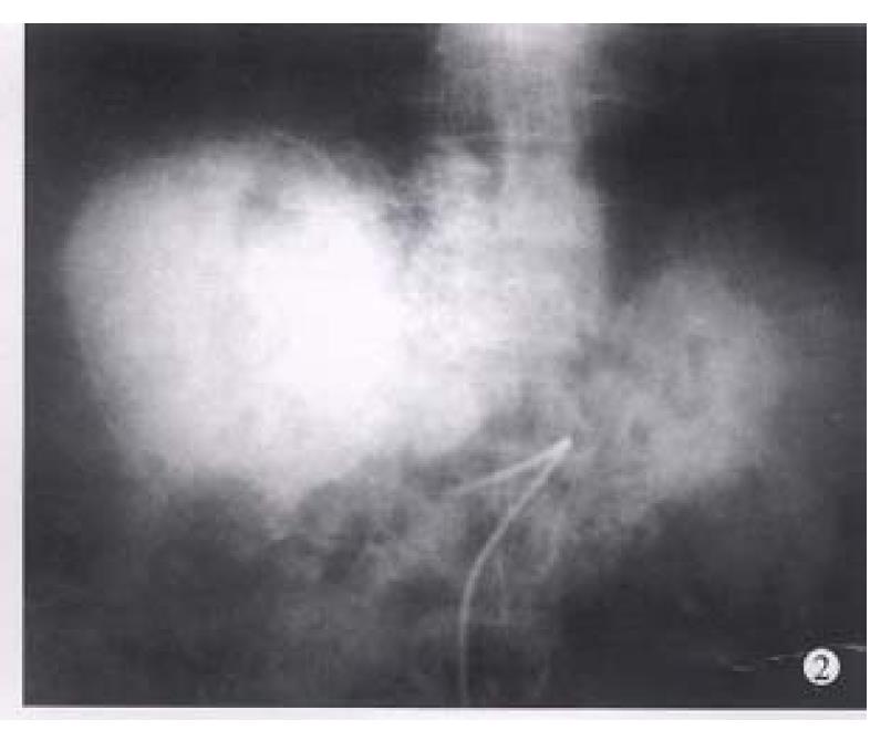
Figure 2 Abdominal plain film immediately after TAE shows accumulation of lipiodol within the tumor.
Figure 3 A parenchymal phase of the hepatic arteriography.
Tumor mass staining is well visualized.
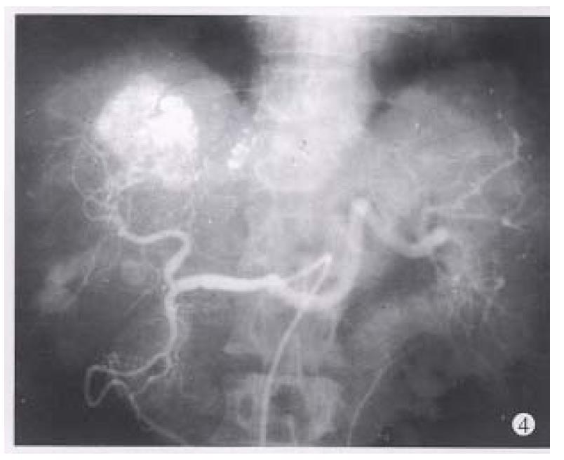
Figure 4 The hepatic angiography after three times of TAE shows occluded branches of right hepatic artery with some collateral vessels and remarkable tumor reduction.
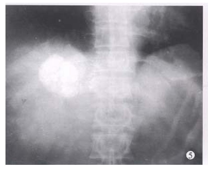
Figure 5 Abdominal plain film after four times of TAE shows deposition of lipiodol within the tumor.
The diameter of tumor was reduced to 4 cm.
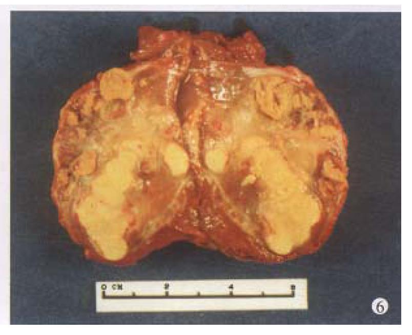
Figure 6 After four times of TAE, the tumor was resected.
The specimen showed necrosis of tumor and fibrotic capsule.
- Citation: Wang JH, Lin G, Yan ZP, Wang XL, Cheng JM, Li MQ. Stage II surgical resection of hepatocellular carcinoma after TAE: a report of 38 cases. World J Gastroenterol 1998; 4(2): 133-136
- URL: https://www.wjgnet.com/1007-9327/full/v4/i2/133.htm
- DOI: https://dx.doi.org/10.3748/wjg.v4.i2.133


