
















Copyright: ©Author(s) 2026.
World J Gastroenterol. Oct 28, 2026; 32(40): 120622
Published online Oct 28, 2026. doi: 10.3748/wjg.120622
Published online Oct 28, 2026. doi: 10.3748/wjg.120622
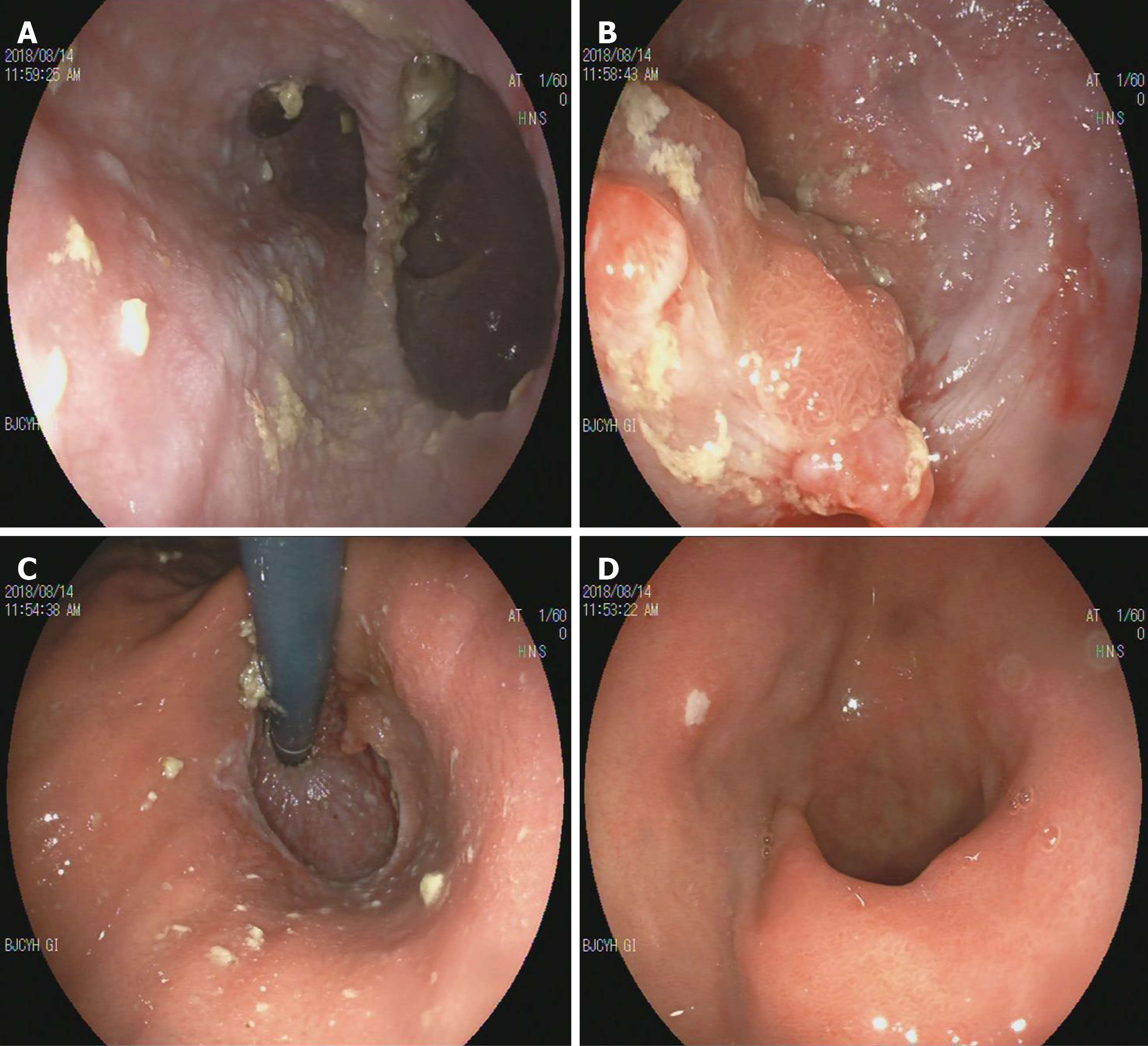
Figure 1 The endoscopic image of patient.
A: A large diverticulum of 6 cm in diameter was seen at the esophagus (30 cm from the incisors), with a large amount of food retention; B: Two polypoid-like bulges in the proximal dentate line, about 0.4-0.8 cm in size; C: The mucosa was smooth and the hernia sac was visible; D: The 0.4 cm shallow ulcer was seen on the anterior wall of the duodenal bulb.
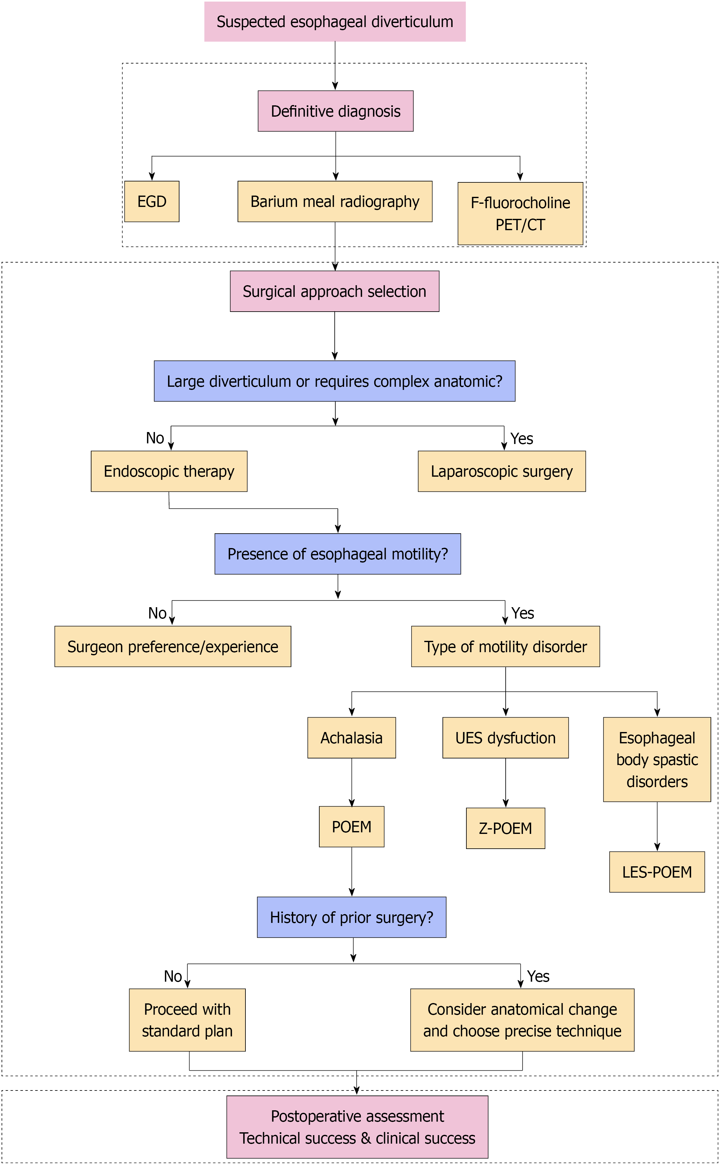
Figure 2 Algorithm for the endoscopic management of esophageal diverticula.
EGD: Esophagogastroduodenoscopy; PET/CT: Positron emission tomography/computed tomography; POEM: Peroral endoscopic myotomy; LES-POEM: Lower esophageal sphincter-sparing peroral endoscopic myotomy.
- Citation: Song XR, Liu YX, Wu XF, Xue R. Esophageal diverticulum: A clinical framework for patient-tailored endoscopic management. World J Gastroenterol 2026; 32(40): 120622
- URL: https://www.wjgnet.com/1007-9327/full/v32/i40/120622.htm
- DOI: https://dx.doi.org/10.3748/wjg.120622