
















Copyright: ©Author(s) 2026.
World J Gastroenterol. Sep 28, 2026; 32(36): 118689
Published online Sep 28, 2026. doi: 10.3748/wjg.118689
Published online Sep 28, 2026. doi: 10.3748/wjg.118689
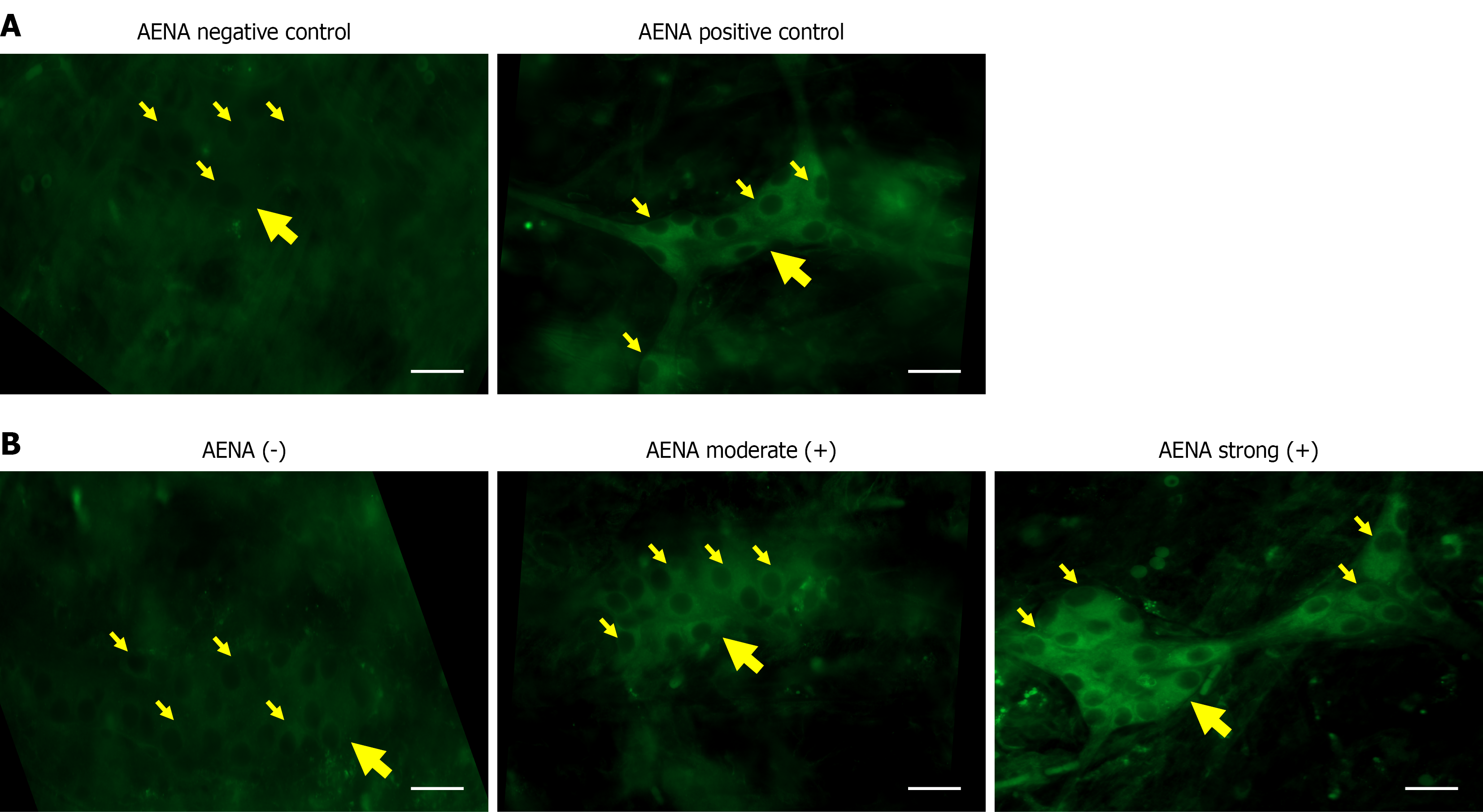
Figure 1 Immunofluorescence staining patterns of anti-enteric neuronal antibodies.
A: Indirect immunofluorescence images of anti-enteric neuronal antibody (AENA)-positive and negative controls; B: Representative indirect immunofluorescence images demonstrating AENA negative (-), moderate-positive, and strong-positive staining intensities. Immunostaining was predominantly localized to the cytoplasm. Large yellow arrows indicate submucosal ganglia, and small yellow arrows indicate myenteric neurons (scale bar = 20 μm). AENA: Anti-enteric neuronal antibody.
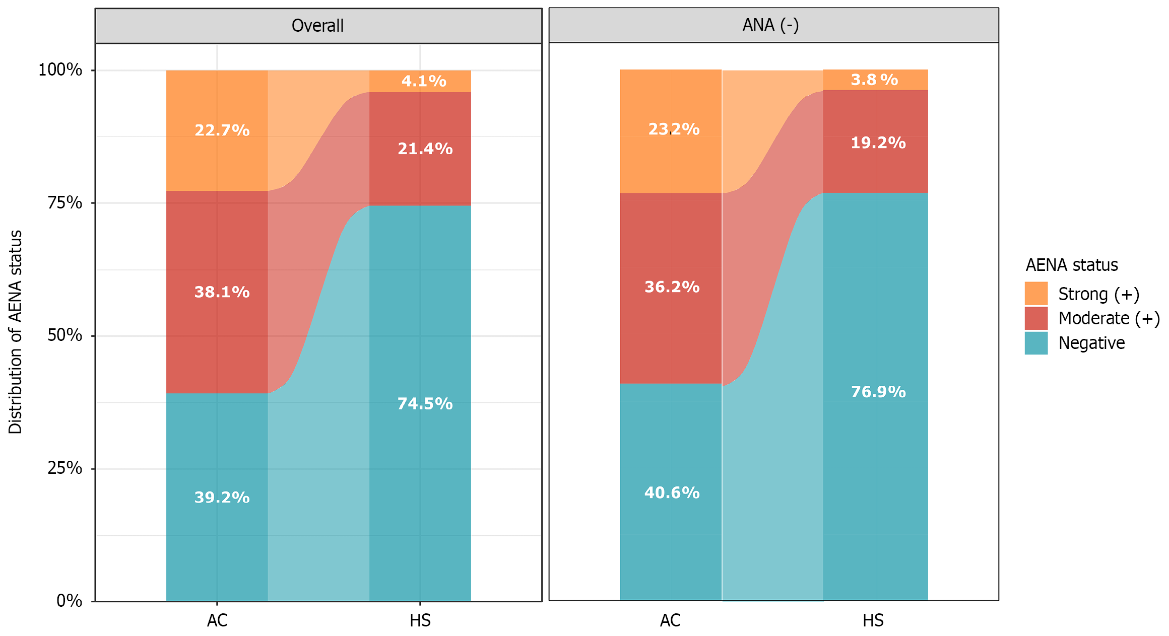
Figure 2 Distribution and overlap of serum anti-enteric neuronal antibodies and antinuclear antibodies in patients with achalasia.
The stacked bar chart displays the distribution of anti-enteric neuronal antibodies (AENA) status stratified by antinuclear antibodies (ANA) classification. Among ANA-negative patients, 59.4% were AENA-positive and 40.6% were AENA-negative, compared with 60.8% and 39.2%, respectively, in the overall achalasia cohort. AENA: Anti-enteric neuronal antibody; ANA: Antinuclear antibodies; AC: Achalasia of cardia; HS: Healthy subjects.
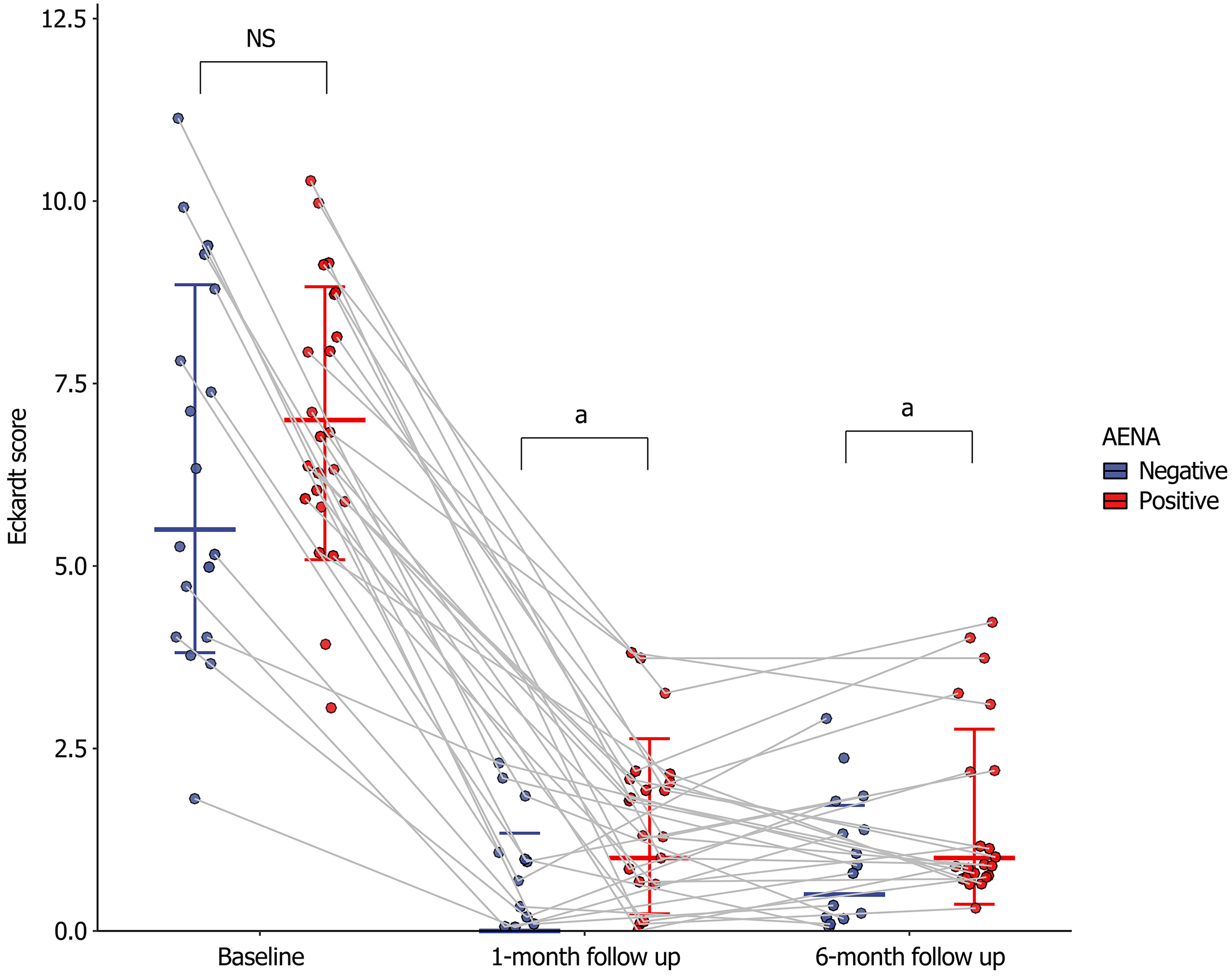
Figure 3 Longitudinal changes in Eckardt scores before and after peroral endoscopic myotomy stratified by anti-enteric neuronal antibody status.
Individual patient trajectories are depicted by gray lines. Scatter plots represent anti-enteric neuronal antibodies (AENA)-negative (blue) and AENA-positive (red) groups. Horizontal and vertical bars indicate median values with interquartile ranges. Eckardt scores were significantly higher in the AENA-positive group at 1-month and 6-month follow-up (P < 0.05). aP < 0.05. NS: Not significant; AENA: Anti-enteric neuronal antibody.
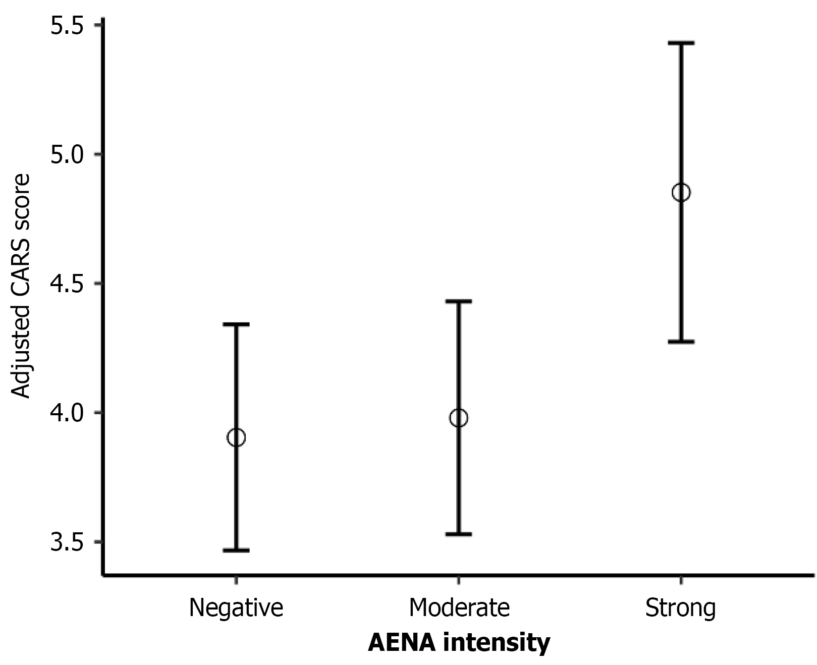
Figure 4 Multivariable linear regression analysis of anti-enteric neuronal antibody intensity and endoscopic severity using the novel endoscopic scoring for achalasia contents, anatomy, resistance, and stasis score.
Adjusted predicted mean CARS (contents, anatomy, resistance, and stasis) scores stratified by anti-enteric neuronal antibody intensity; error bars represent 95% confidence intervals. AENA: Anti-enteric neuronal antibody; CARS: Contents, anatomy, resistance, and stasis.
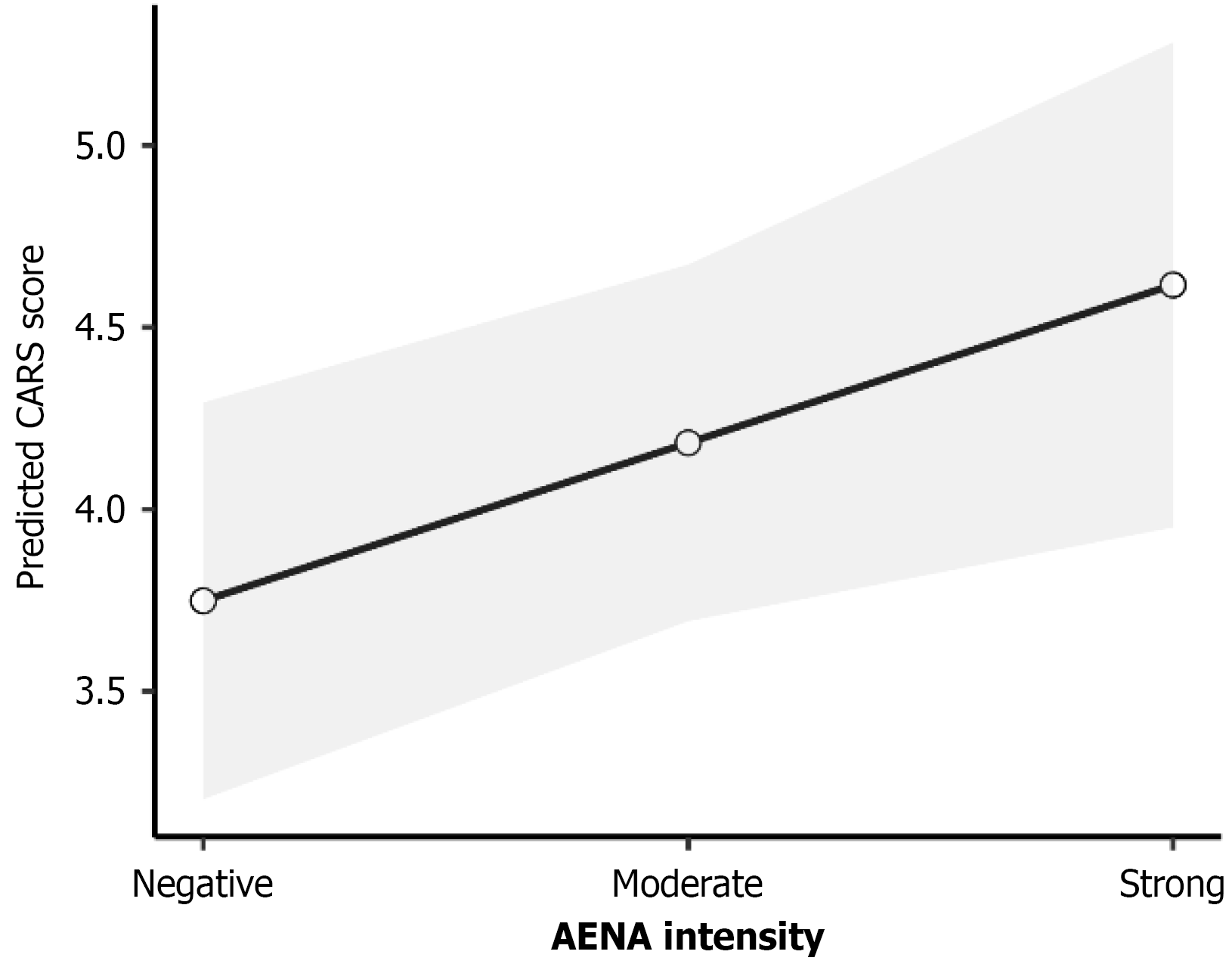
Figure 5 Dose-response association between anti-enteric neuronal antibody intensity and contents, anatomy, resistance, and stasis scores.
Predicted marginal means of CARS (contents, anatomy, resistance, and stasis) scores across anti-enteric neuronal antibody intensity levels. Anti-enteric neuronal antibody intensity was analyzed as an ordinal variable (negative = 0; moderate-positive = 1; strong-positive = 2). The solid line indicates the linear trend, and the shaded area represents the 95% confidence interval. AENA: Anti-enteric neuronal antibody; CARS: Contents, anatomy, resistance, and stasis.
- Citation: Hu YW, Tang P, Guo T, Wang ZF, Fang XC, Zhu LM, Fei GJ, Chen Y, Li XQ. Serum anti-enteric neuronal antibodies in patients with achalasia and their association with clinical profiles. World J Gastroenterol 2026; 32(36): 118689
- URL: https://www.wjgnet.com/1007-9327/full/v32/i36/118689.htm
- DOI: https://dx.doi.org/10.3748/wjg.118689