
















Copyright: ©Author(s) 2026.
World J Gastroenterol. Sep 28, 2026; 32(36): 118689
Published online Sep 28, 2026. doi: 10.3748/wjg.118689
Published online Sep 28, 2026. doi: 10.3748/wjg.118689
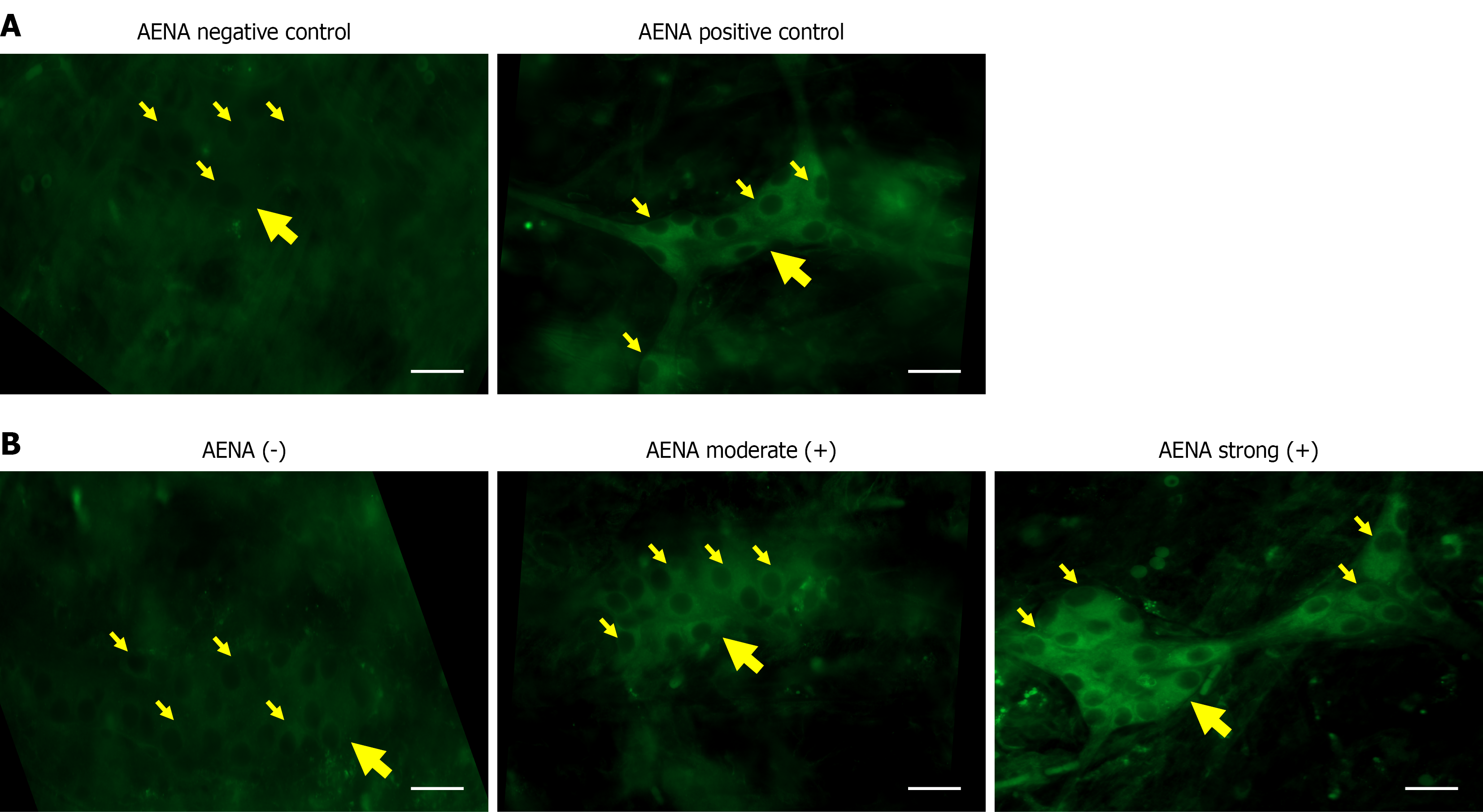
Figure 1 Immunofluorescence staining patterns of anti-enteric neuronal antibodies.
A: Indirect immunofluorescence images of anti-enteric neuronal antibody (AENA)-positive and negative controls; B: Representative indirect immunofluorescence images demonstrating AENA negative (-), moderate-positive, and strong-positive staining intensities. Immunostaining was predominantly localized to the cytoplasm. Large yellow arrows indicate submucosal ganglia, and small yellow arrows indicate myenteric neurons (scale bar = 20 μm). AENA: Anti-enteric neuronal antibody.
- Citation: Hu YW, Tang P, Guo T, Wang ZF, Fang XC, Zhu LM, Fei GJ, Chen Y, Li XQ. Serum anti-enteric neuronal antibodies in patients with achalasia and their association with clinical profiles. World J Gastroenterol 2026; 32(36): 118689
- URL: https://www.wjgnet.com/1007-9327/full/v32/i36/118689.htm
- DOI: https://dx.doi.org/10.3748/wjg.118689