Copyright
©The Author(s) 2025.
World J Gastroenterol. May 14, 2025; 31(18): 105443
Published online May 14, 2025. doi: 10.3748/wjg.v31.i18.105443
Published online May 14, 2025. doi: 10.3748/wjg.v31.i18.105443
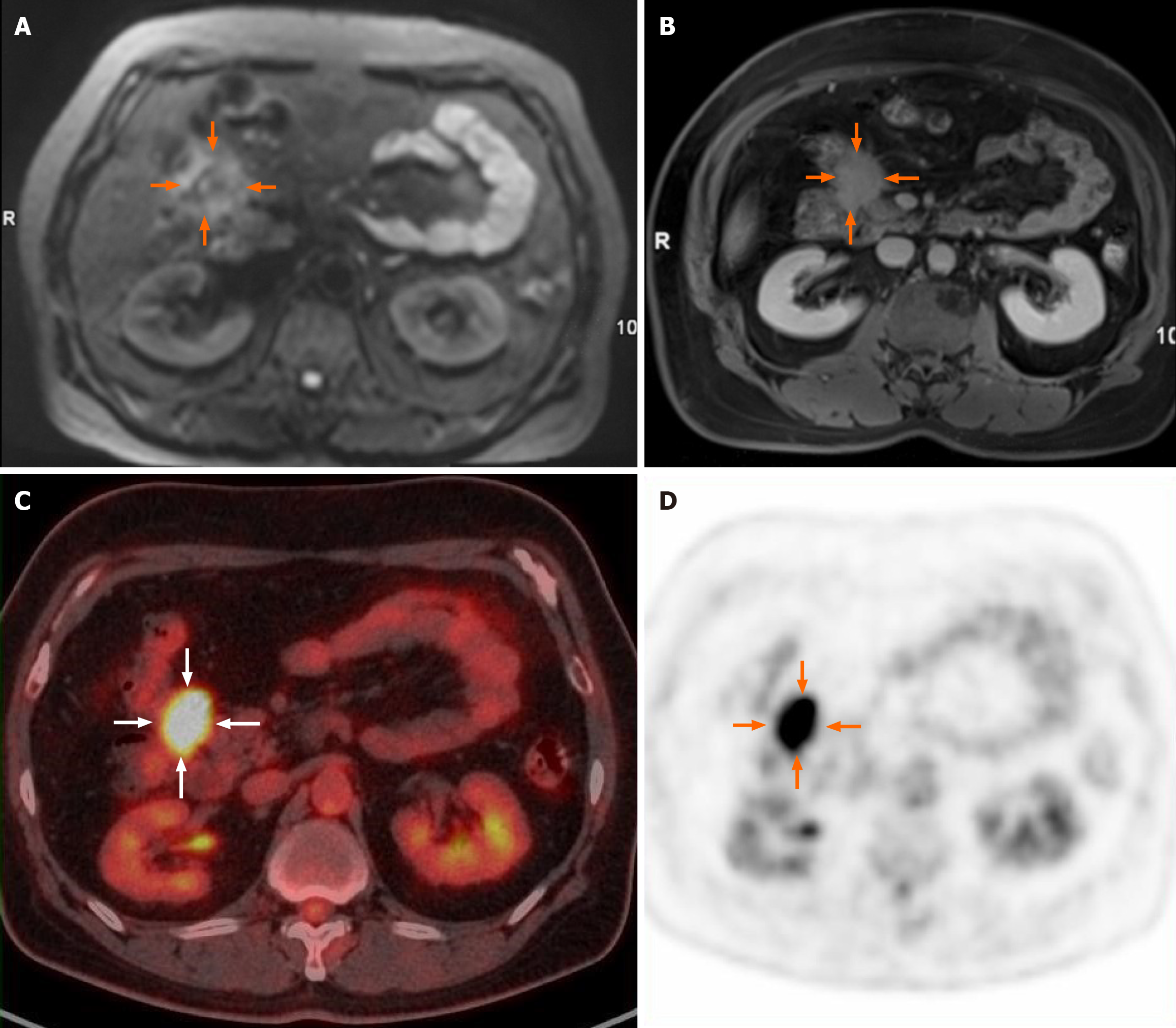
Figure 1 Magnetic resonance imaging and 18F-fluorodeoxyglucose positron emission tomography images.
A and B: Diffusion-weighted imaging (A) and delayed-enhancement (B) images of magnetic resonance imaging showed an irregular soft tissue with ill-defined boundary with the head of pancreas and the hepatic flexure of the colon; C and D: On 18F-fluorodeoxyglucose positron emission tomography images, increased uptake in the tumor with a maximum standardized uptake value of 10.3 was detected.
Figure 2 Pathological result.
A: Idiopathic retroperitoneal fibrosis; B: Lymphoid plasma cell infiltration to the muscularis propria of the colon.
- Citation: Dong ZY, Zhu HB. Idiopathic retroperitoneal fibrosis arising from peritoneal space: A case report and review of literature. World J Gastroenterol 2025; 31(18): 105443
- URL: https://www.wjgnet.com/1007-9327/full/v31/i18/105443.htm
- DOI: https://dx.doi.org/10.3748/wjg.v31.i18.105443


