Copyright
©The Author(s) 2022.
World J Gastroenterol. Oct 7, 2022; 28(37): 5506-5514
Published online Oct 7, 2022. doi: 10.3748/wjg.v28.i37.5506
Published online Oct 7, 2022. doi: 10.3748/wjg.v28.i37.5506
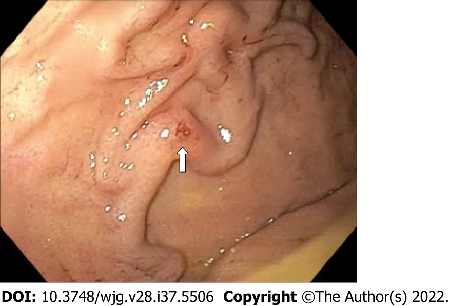
Figure 1 Emergent esophagogastroduodenoscopy: Retroflexed view of the gastric fundus showing varicose-shaped submucosal vessels with a small erosion (arrow).
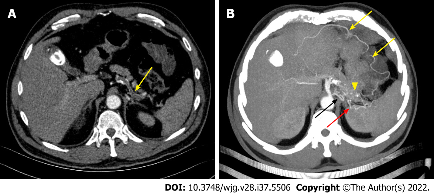
Figure 2 Emergent computed tomography angiography: Axial view.
A: Arterial phase showing complete splenic artery thrombosis (arrow); B: Arterial phase curved-multiplanar reconstruction showing splenic artery collateral vessels (yellow arrows) with an arterial cluster at the gastric fundus (arrowhead) arising from the left gastric artery (black arrow) and the left gastroepiploic artery (red arrow).
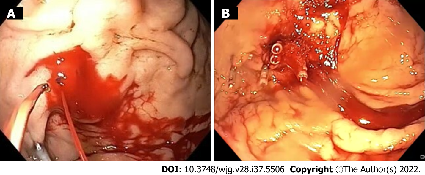
Figure 3 Therapeutic esophagogastroduodenoscopy.
A: Spurting active bleeding of the gastric submucosal arterial collaterals after first endoclip application; B: Successful mechanical hemostasis achievement.
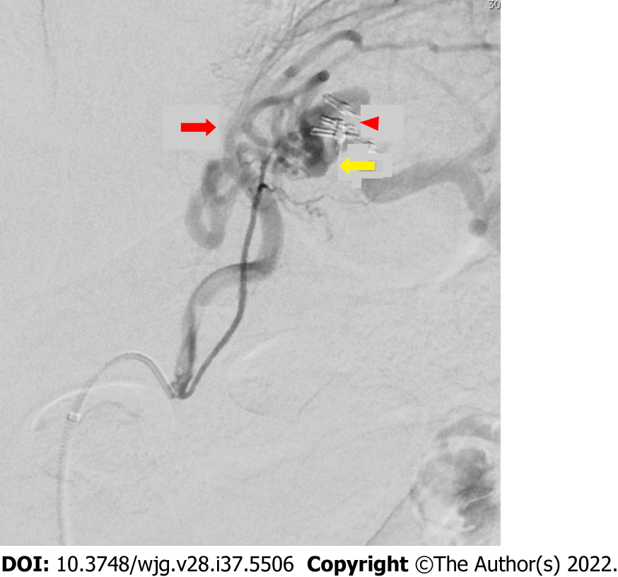
Figure 4 Operative angiography showing a cluster of collateral arterial vessels emerging from the left gastric artery (red arrow) and the left gastroepiploic artery (yellow arrow) projecting into the gastric fundus.
It also shows previously placed endoclips on the submucosal collateral arteries of the gastric fundus without active contrast extravasation (arrowhead).
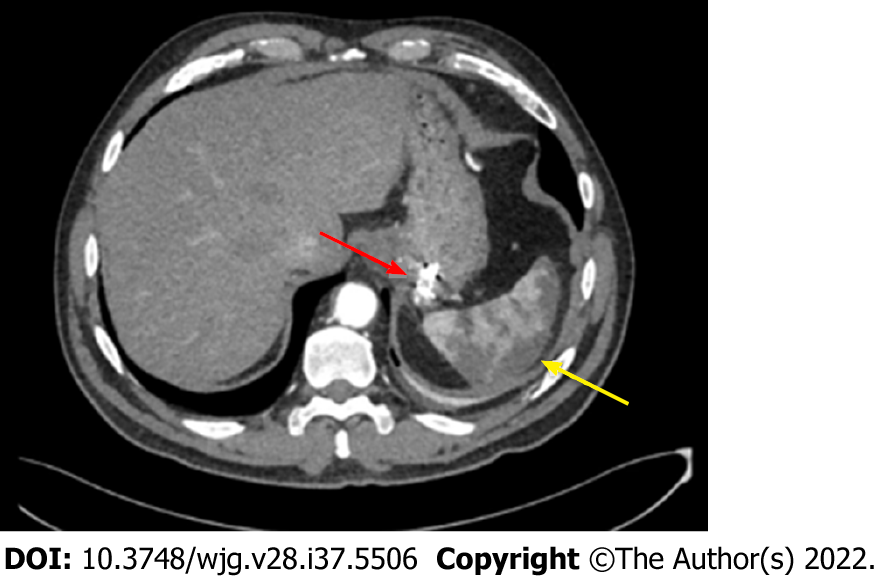
Figure 5 Post-operative computed tomography angiography: Axial view showing collateral arteries of the gastric fundus treated by endoscopic clipping plus microspheres embolization (red arrow) and splenic infarction (yellow arrow).
- Citation: Martino A, Di Serafino M, Zito FP, Maglione F, Bennato R, Orsini L, Iacobelli A, Niola R, Romano L, Lombardi G. Massive bleeding from gastric submucosal arterial collaterals secondary to splenic artery thrombosis: A case report. World J Gastroenterol 2022; 28(37): 5506-5514
- URL: https://www.wjgnet.com/1007-9327/full/v28/i37/5506.htm
- DOI: https://dx.doi.org/10.3748/wjg.v28.i37.5506


