Copyright
©The Author(s) 2022.
World J Gastroenterol. Oct 7, 2022; 28(37): 5457-5468
Published online Oct 7, 2022. doi: 10.3748/wjg.v28.i37.5457
Published online Oct 7, 2022. doi: 10.3748/wjg.v28.i37.5457
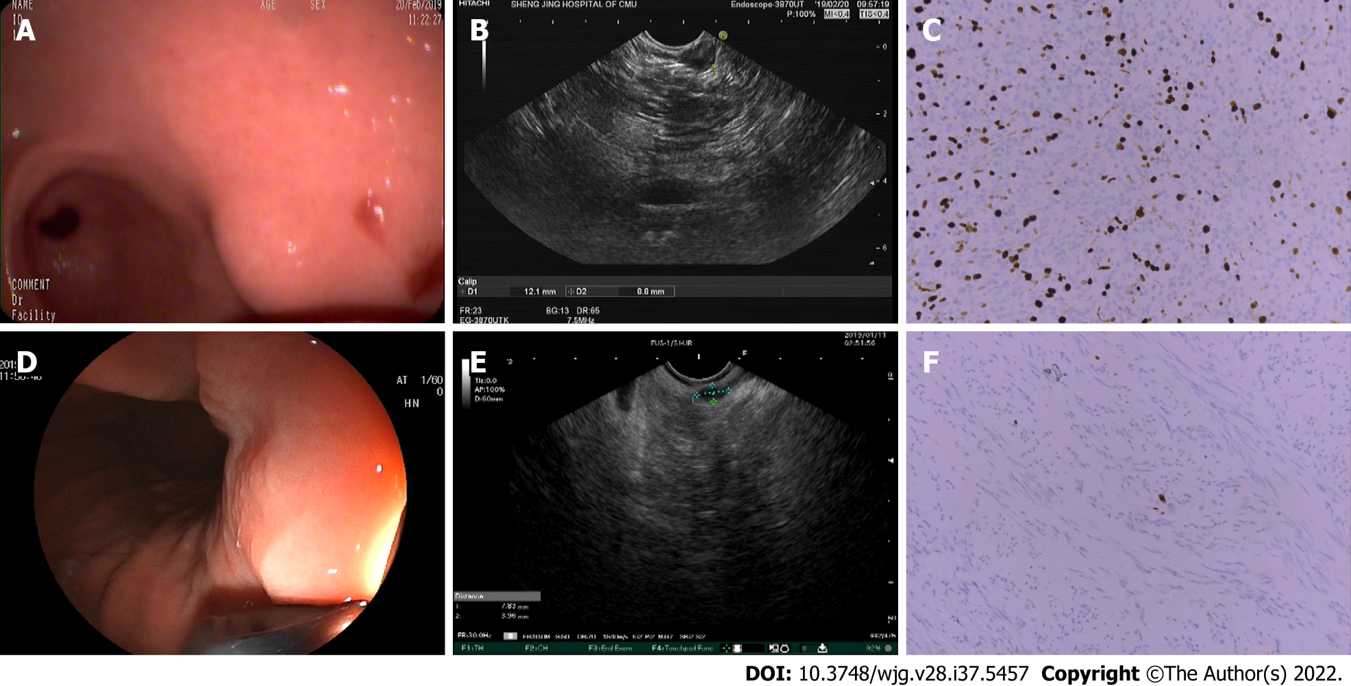
Figure 1 Endoscopic ultrasound imaging features were consistent with the postoperative pathological results.
A: A lesion with a diameter of approximately 1.2 cm was observed in the gastric antrum using an endoscope; B: Heterogeneity was observed as a characteristic of endoscopic ultrasound (EUS) imaging; C: Gastric stromal tumor (intermediate risk). The spindle of the tumor cells is arranged in sheets and bundles with nuclear division of approximately 8-9/50 high power field. Immunohistochemistry: CD117 (+); CD34 (-); Desmin (-); Dog1 (+); Ki-67 (35%+); smooth muscle actin (+); and S-100 (-); D: A lesion with a diameter of approximately 0.6 cm was observed in the gastric fundus using an endoscope; E: EUS images were characterized by a hypoechoic mass in the muscularis propria; F: Gastric stromal tumor (very low risk). The spindle of the tumor cells is arranged in spindle and bundles without obvious nuclear division. Immunohistochemistry: CD117 (+); CD34 (-); Desmin (-); Dog1 (+); Ki-67 (< 1%+); smooth muscle actin (-); and S-100 (-).
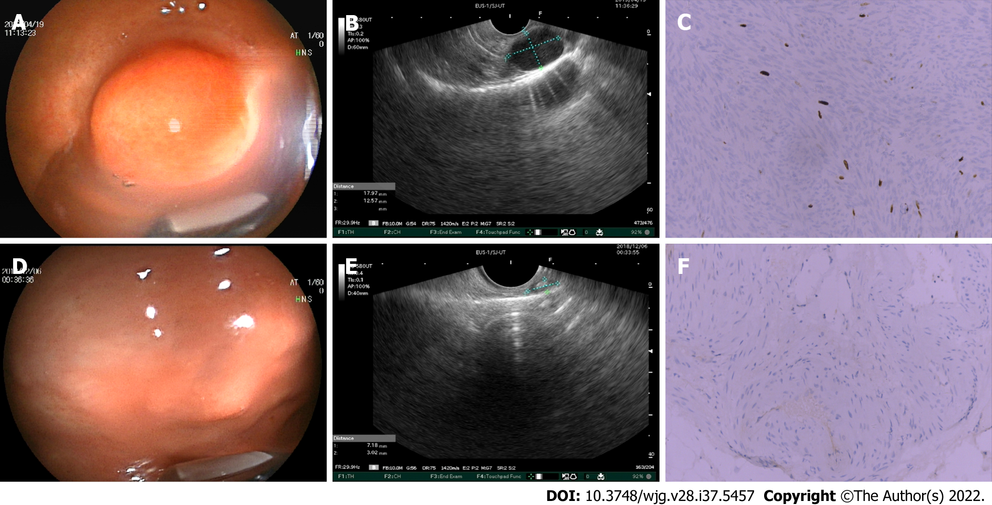
Figure 2 Endoscopic ultrasound imaging features were inconsistent with the postoperative pathological results.
A: A lesion with a diameter of approximately 2 cm was observed in the gastric fundus using an endoscope; B: Endoscopic ultrasound (EUS) images were characterized by a hypoechoic mass in the muscularis propria; C: Gastric stromal tumor (intermediate risk). The spindle of the tumor cells is arranged in a spindle and braided pattern with nuclear division of approximately 8/50 high power field. Immunohistochemistry: CD117 (+); CD34 (-); Desmin (-); Dog1 (+); Ki-67 (10%+); smooth muscle actin (-); and S-100 (-); D: A lesion with a diameter of approximately 0.6 cm was observed in the gastric fundus using an endoscope; E: Strong echogenic foci and heterogeneity were observed as characteristics of EUS imaging; F: Gastric stromal tumor (very low risk). The spindle of the tumor cells is arranged in spindle and bundles without obvious nuclear division. Some tissues calcify. Immunohistochemistry: CD117 (+); CD34 (+); Desmin (-); Ki-67 (1%+); smooth muscle actin (+); and S-100 (-).
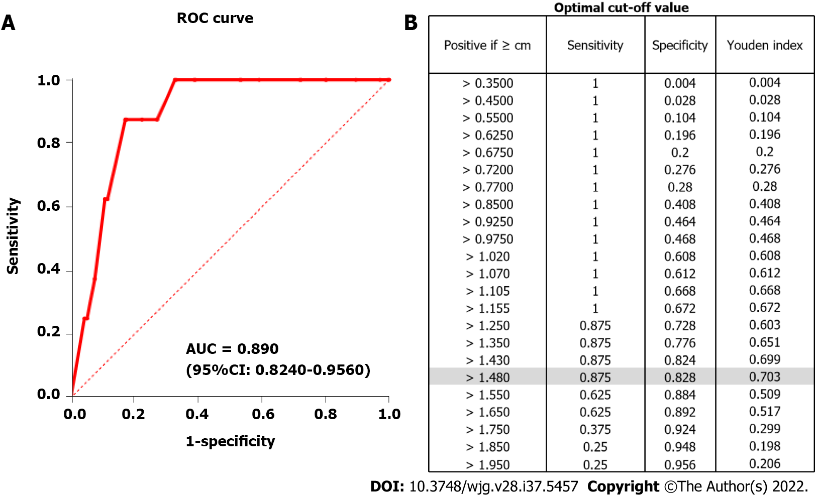
Figure 3 Receiver operating characteristic curve.
A: The receiver operating characteristic curve of the cut-off (1.48 cm); B: The optimal cut-off (1.48 cm) with a sensitivity of 0.875 and a specificity of 0.828. ROC: Receiver operating characteristic; AUC: Area under the curve.
- Citation: Ge QC, Wu YF, Liu ZM, Wang Z, Wang S, Liu X, Ge N, Guo JT, Sun SY. Efficacy of endoscopic ultrasound in the evaluation of small gastrointestinal stromal tumors. World J Gastroenterol 2022; 28(37): 5457-5468
- URL: https://www.wjgnet.com/1007-9327/full/v28/i37/5457.htm
- DOI: https://dx.doi.org/10.3748/wjg.v28.i37.5457


