Copyright
©The Author(s) 2022.
World J Gastroenterol. Aug 7, 2022; 28(29): 3838-3853
Published online Aug 7, 2022. doi: 10.3748/wjg.v28.i29.3838
Published online Aug 7, 2022. doi: 10.3748/wjg.v28.i29.3838
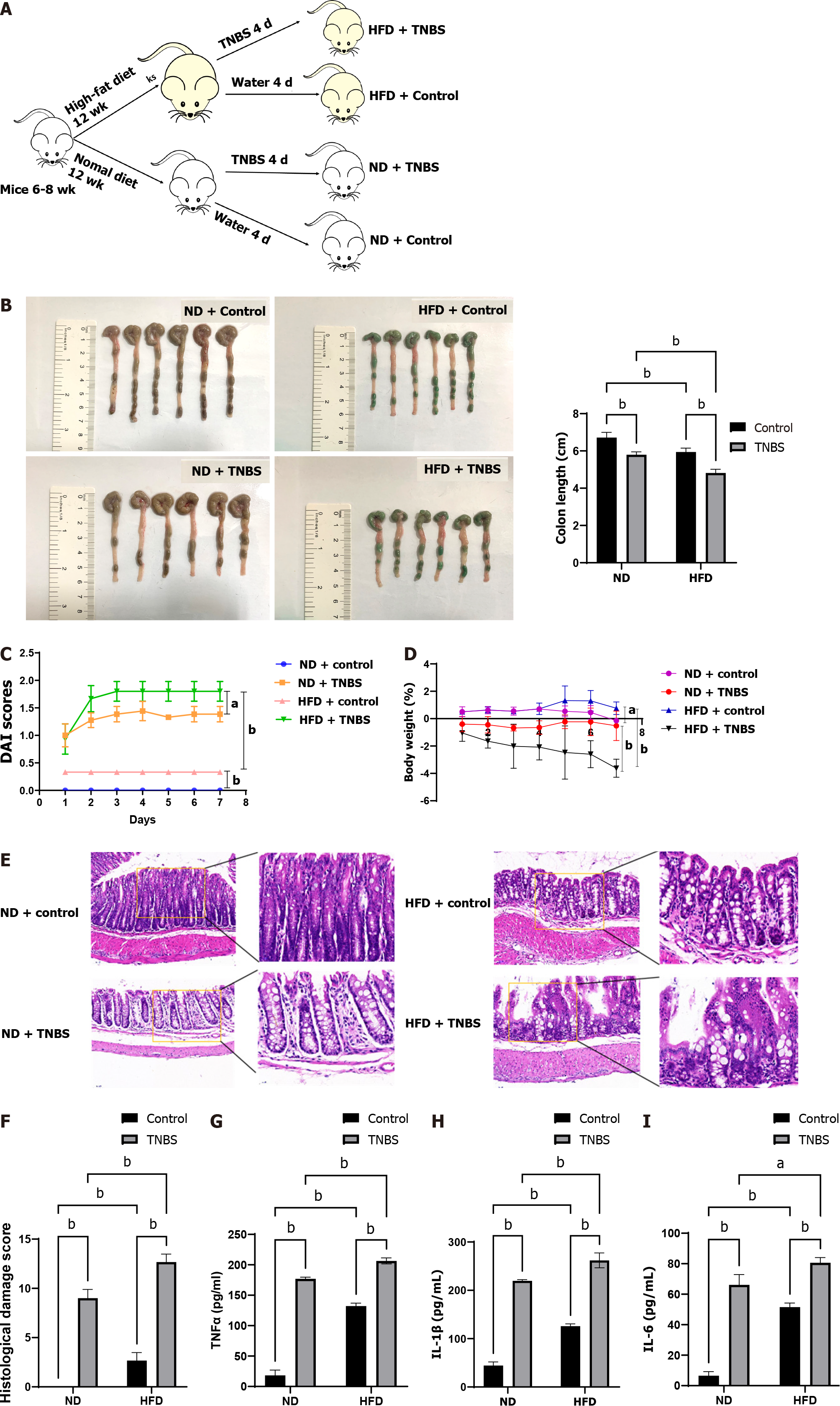
Figure 1 High-fat diet aggravated colitis.
A: Experimental grouping process; B: Colon length of mice in each group; C: Disease activity index of mice in each group; D: Body weight change trend of mice in each group; E: Hematoxylin and eosin staining images of colon tissue; F: Histological damage score of colon tissue; G: Expression of tumor necrosis factor-α in colon tissue by enzyme linked immunosorbent assay (ELISA); H: Expression of interleukin-1β in colon tissue by ELISA; I: Expression of interleukin-6 (IL-6) in colon tissue by ELISA. DAI: Disease activity index; TNBS: 2,4,6-trinitrobe-nzenesulfonic acid; HFD: High-fat diet; ND: Normal diet; TNF-α: Tumor necrosis factor-α; IL-1β: Interleukin-1β. aP < 0.05, bP < 0.01.
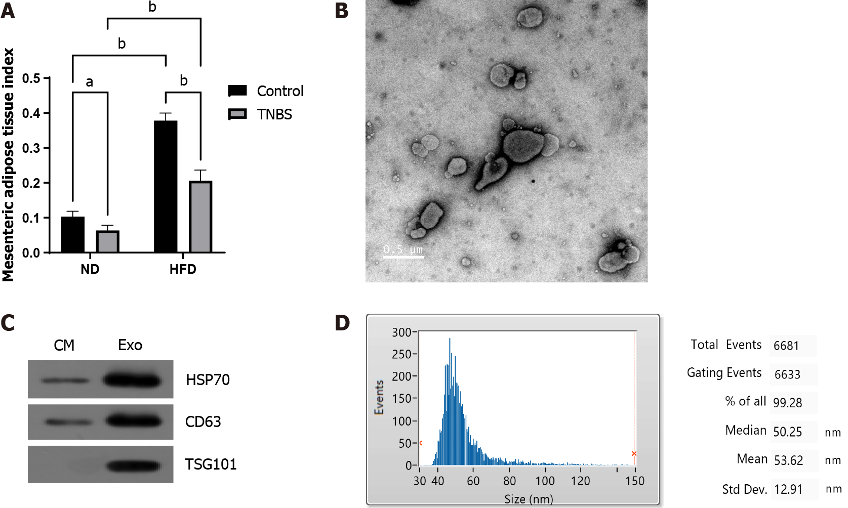
Figure 2 Extraction and identification of exosomes derived from mesenteric adipose tissue.
A: Mesenteric adipose index of mice in each group; B: Images of exosomes observed under transmission electron microscopy; C: Western blotting to detect exosome marker proteins; D: Exosome particle size analysis. TNBS: 2,4,6-trinitrobe-nzenesulfonic acid; HFD: High-fat diet; ND: Normal diet. aP < 0.05, bP < 0.01.
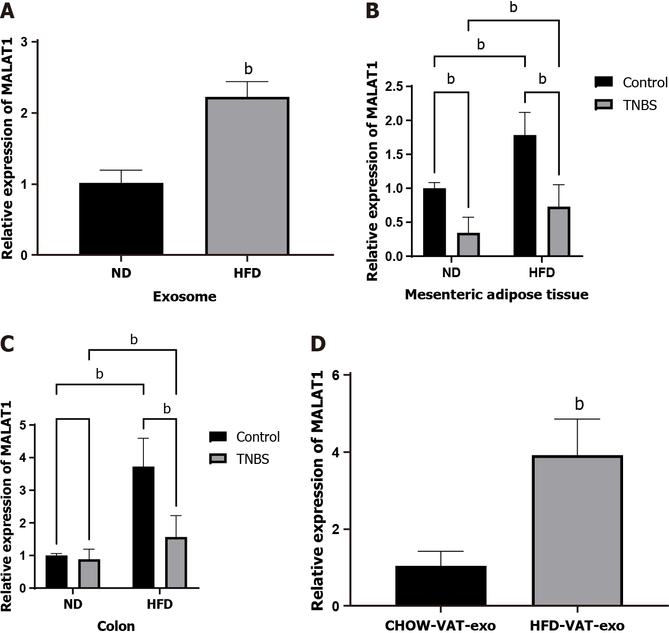
Figure 3 Mesenteric adipose tissue derived exosomes from high-fat diet-fed mice increase the expression of long noncoding RNAs metastasis-associated lung adenocarcinoma transcript 1 in colon.
A: Expression of metastasis-associated lung adenocarcinoma transcript 1 (MALAT1) in mesenteric adipose tissue derived exosomes (MAT-Exos) in each group; B: Expression of MALAT1 in mesenteric adipose tissue in each group; C: Expression of MALAT1 in colon in each group; D: Expression of MALAT1 in colon of normal mice after treated with MAT-Exos extracted from higt-fat diet mice or normal diet mice. MALAT1: Metastasis-associated lung adenocarcinoma transcript 1; TNBS: 2,4,6-trinitrobe-nzenesulfonic acid; HFD: High-fat diet; ND: Normal diet. bP < 0.01.
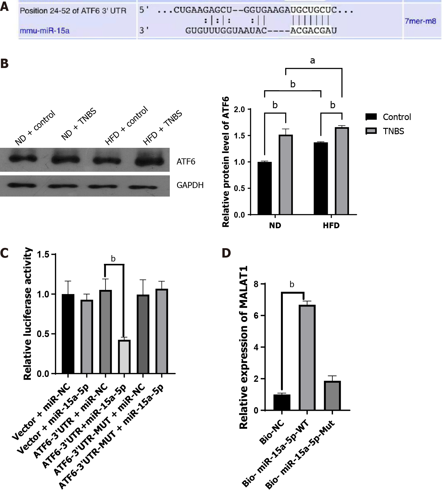
Figure 4 Associations between metastasis-associated lung adenocarcinoma transcript 1 and miR-15a-5p/activating transcription factor 6.
A: Prediction of miR-15a-5p binding site on the 3’ untranslated region of activating transcription factor 6 (ATF6); B: Western blotting to detect ATF6 protein; C: Dual luciferase report of miR-15a-5p and ATF6; D: RNA pull down analysis of miR-15a-5p and metastasis-associated lung adenocarcinoma transcript 1. ATF6: Activating transcription factor 6; TNBS: 2,4,6-trinitrobe-nzenesulfonic acid; HFD: High-fat diet; ND: Normal diet; 3’ UTR: 3’ untranslated region; WT: Wild type; Mut: Mutant. aP < 0.05, bP < 0.01.
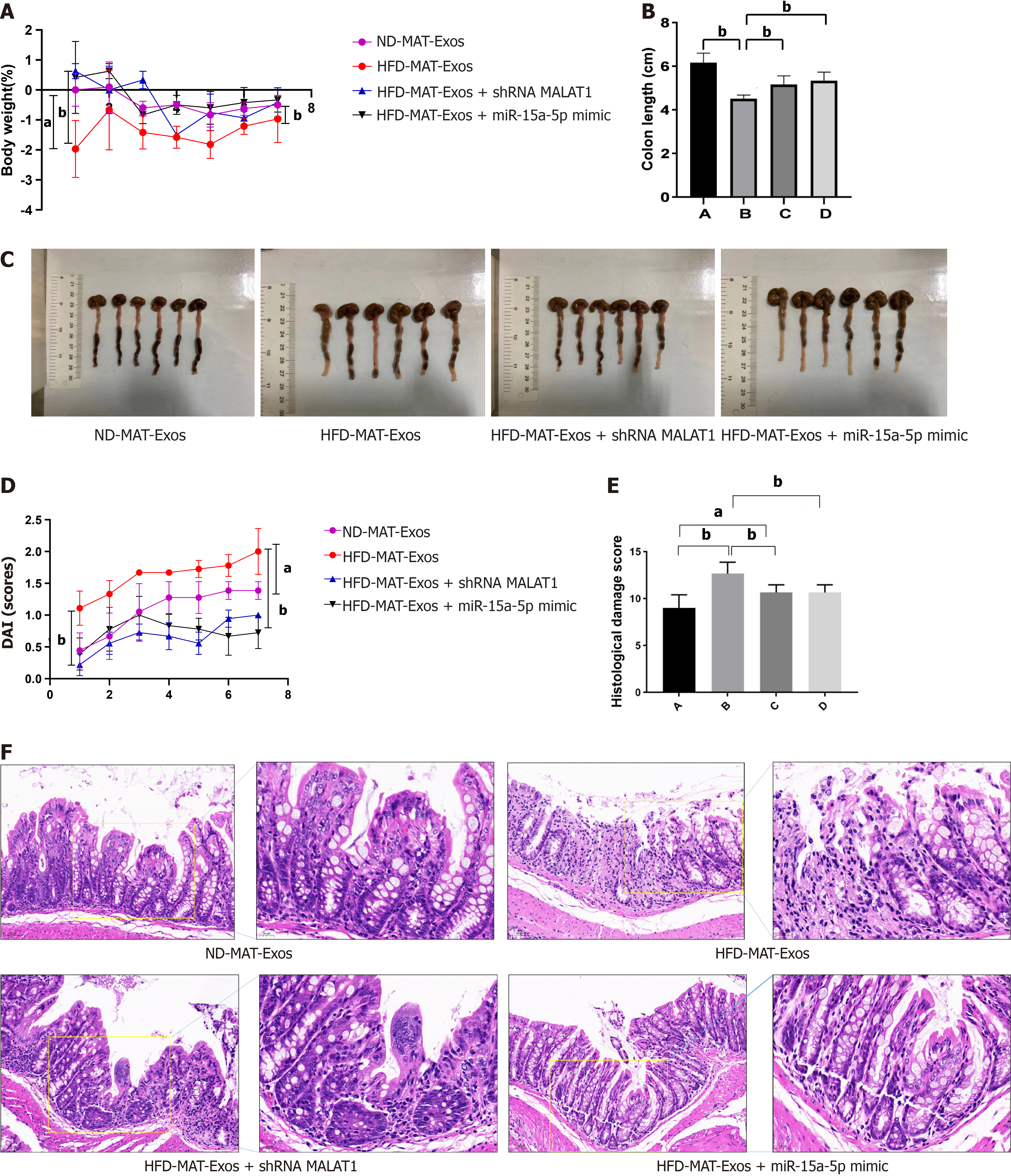
Figure 5 Mesenteric adipose tissue derived exosomes of high-fat diet-fed mice aggravate 2,4,6-trinitrobe-nzenesulfonic acid solution-induced colitis, reversed by shMALAT1 and miR-15a-5p mimic.
A: Body weight change trend of mice in each group; B: Colon length of mice in each group; ND-MAT-Exos, HFD-MAT-Exos, HFD-MAT-Exos + shMALAT1, and HFD-MAT-Exos + miR-15a-5p mimic are labelled as A, B, C, and D groups, respectively; C: Colon image of mice in each group; D: Disease activity index of mice in each group; E: Histological damage score of colon tissue, ND-MAT-Exos, HFD-MAT-Exos, HFD-MAT-Exos + shMALAT1, and HFD-MAT-Exos + miR-15a-5p mimic are labelled as A, B, C, and D groups, respectively; F: Hematoxylin and eosin staining images of colon tissue. HFD: High-fat diet; ND: Normal diet; MAT-Exos: Mesenteric adipose tissue derived exosomes; MALAT1: Metastasis-associated lung adenocarcinoma transcript 1. aP < 0.05, bP < 0.01.
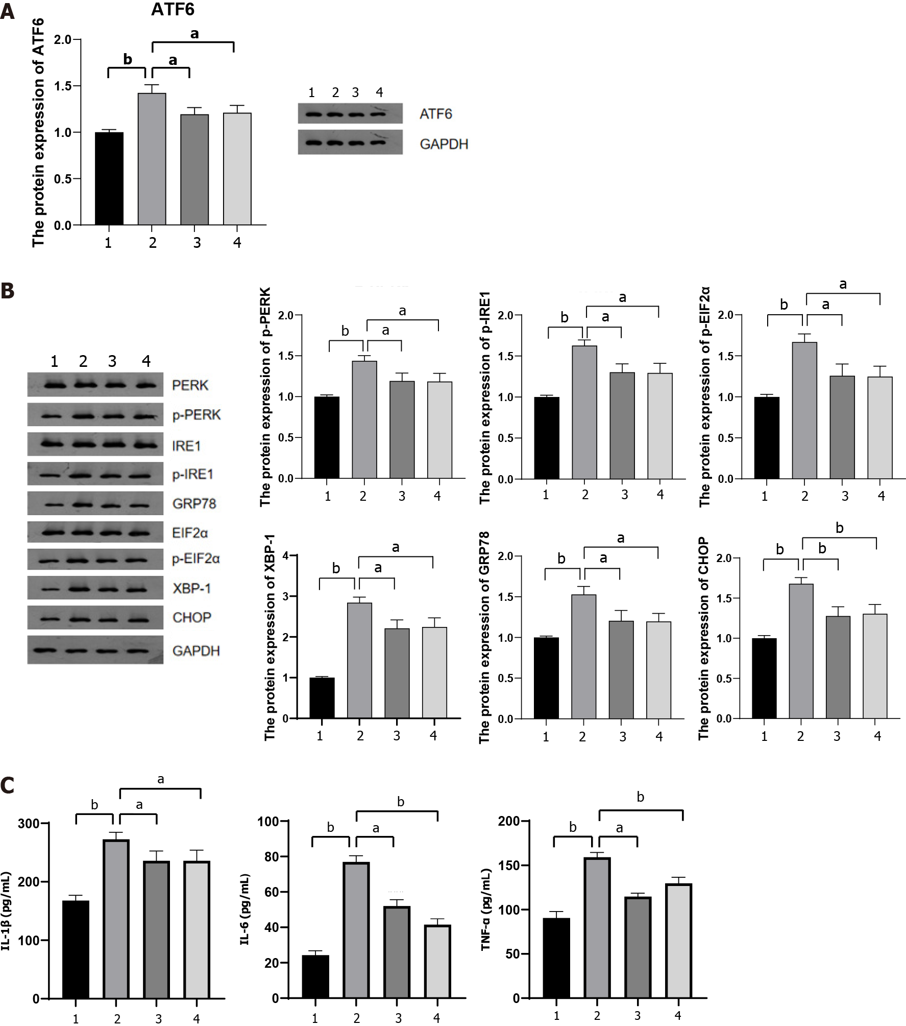
Figure 6 Long noncoding RNAs metastasis-associated lung adenocarcinoma transcript 1 acted on the miR-15a-5p/activating tran
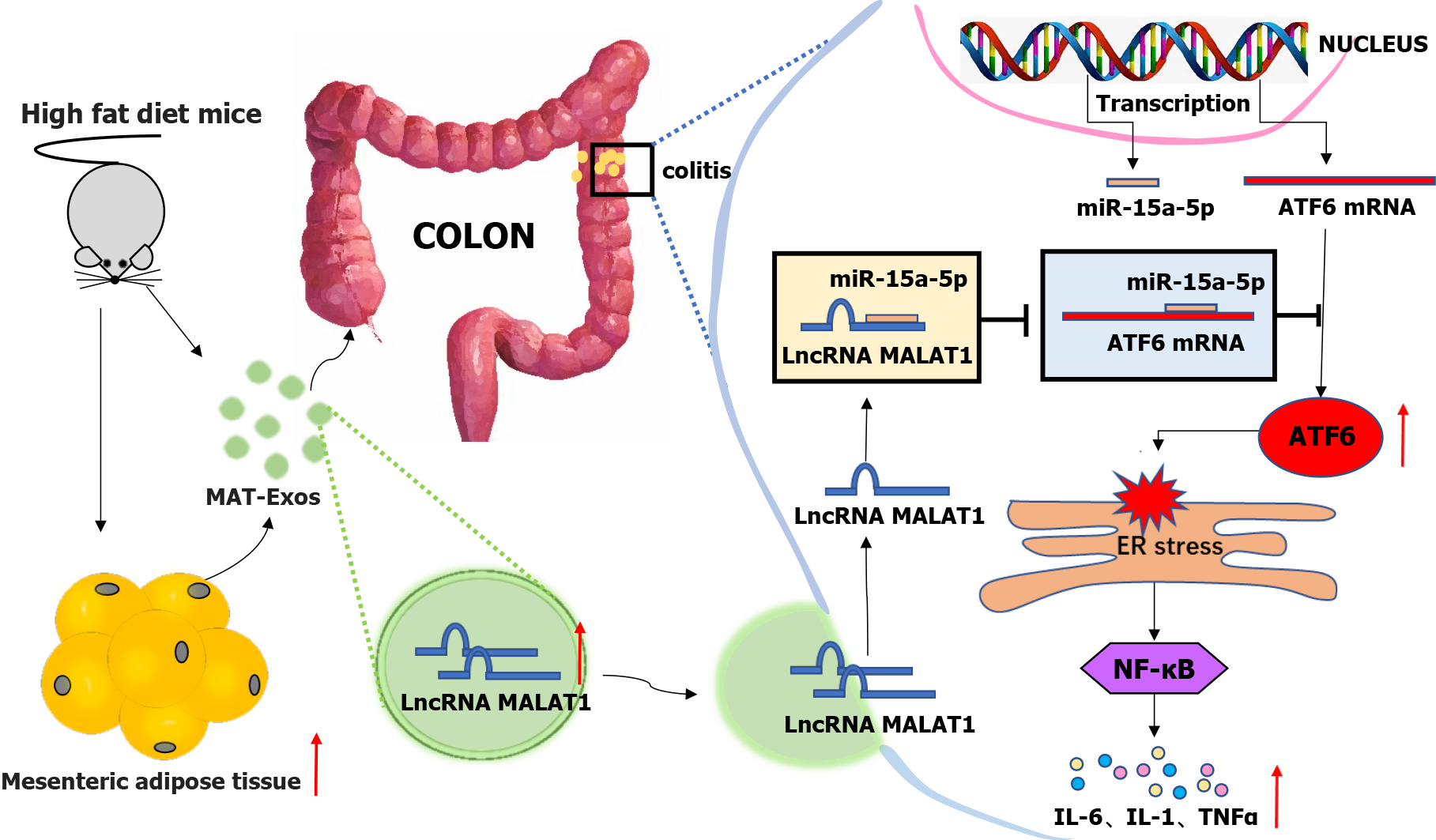
Figure 7 Long non-coding RNA metastasis-associated lung adenocarcinoma transcript 1 encapsulated by mesenteric adipose tissue-derived exosomes targets the colon to aggravate colitis via the microRNA-15a-5p/activating transcription factor 6 axis in response to endoplasmic reticulum stress.
ATF-6: Activating transcription factor 6; LnRNA: Long non-coding RNA; IL: Interleukin; TNF-α: Tumor necrosis factor-α; ER: Endoplasmic reticulum; MAT-Exos: Mesenteric adipose tissue derived exosomes; MALAT1: Metastasis-associated lung adenocarcinoma transcript 1.
- Citation: Chen D, Lu MM, Wang JH, Ren Y, Xu LL, Cheng WX, Wang SS, Li XL, Cheng XF, Gao JG, Kalyani FS, Jin X. High-fat diet aggravates colitis via mesenteric adipose tissue derived exosome metastasis-associated lung adenocarcinoma transcript 1. World J Gastroenterol 2022; 28(29): 3838-3853
- URL: https://www.wjgnet.com/1007-9327/full/v28/i29/3838.htm
- DOI: https://dx.doi.org/10.3748/wjg.v28.i29.3838


