Copyright
©The Author(s) 2021.
World J Gastroenterol. Jan 14, 2021; 27(2): 152-161
Published online Jan 14, 2021. doi: 10.3748/wjg.v27.i2.152
Published online Jan 14, 2021. doi: 10.3748/wjg.v27.i2.152
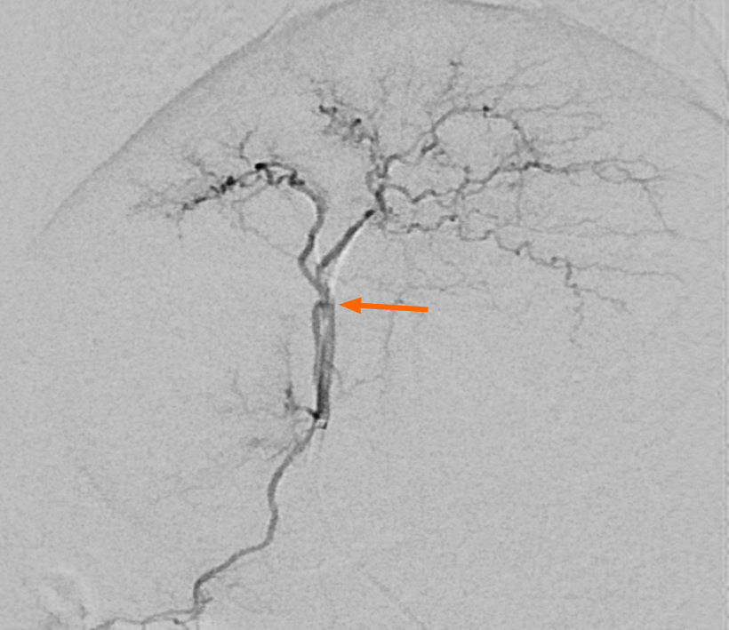
Figure 1 Angiographic findings of the hepatic artery in the rabbit.
The hepatic angiogram shows both lobar hepatic arteries. The catheter tip is located in the proper hepatic artery and passes through the gastroduodenal artery. Triolein emulsion or normal saline, doxorubicin, and evans blue were injected through a microcatheter at this level.
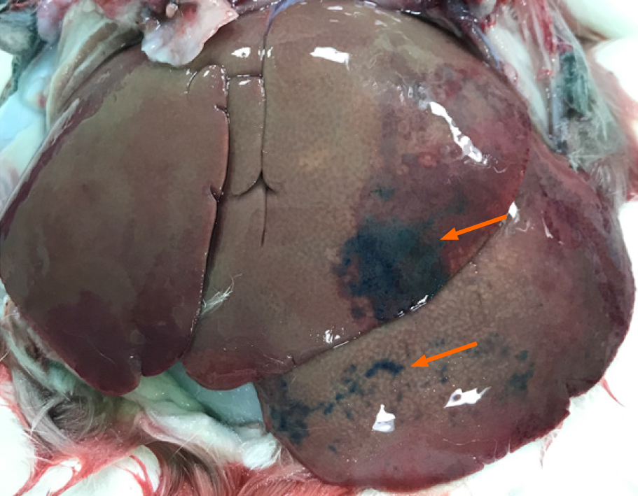
Figure 2 Gross finding of the hepatic surface from a rabbit in the triolein emulsion group after evans blue staining.
The anterior surface of the left lobe shows patchy blue staining by evans blue.
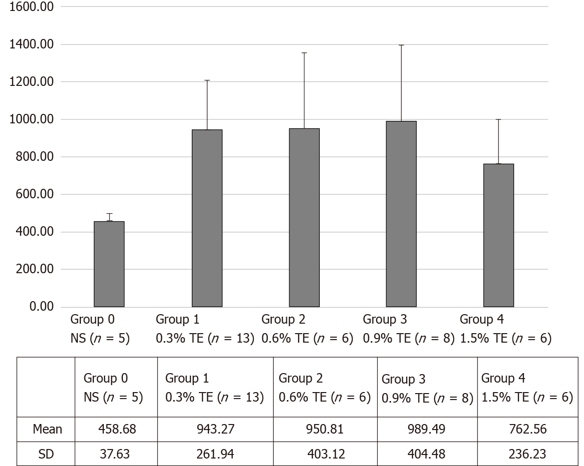
Figure 3 Longitudinal plot of mean doxorubicin concentration in the liver in the control and triolein emulsion groups.
NS: Normal saline; TE: Triolein emulsion.
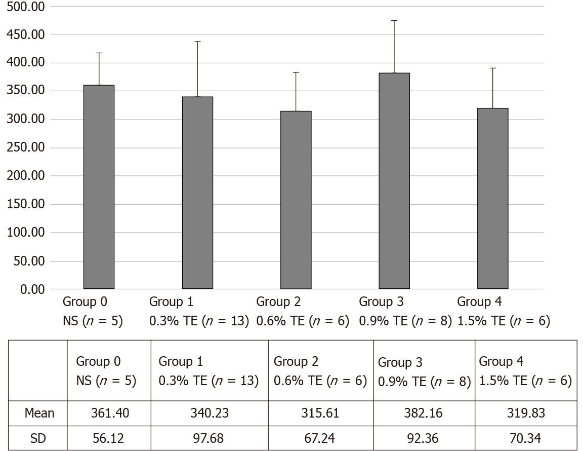
Figure 4 Longitudinal plot of mean doxorubicin concentration in the lungs in the control and triolein emulsion groups.
NS: Normal saline; TE: Triolein emulsion.
- Citation: Kim YW, Kim HJ, Cho BM, Choi SH. Triolein emulsion infusion into the hepatic artery increases vascular permeability to doxorubicin in rabbit liver. World J Gastroenterol 2021; 27(2): 152-161
- URL: https://www.wjgnet.com/1007-9327/full/v27/i2/152.htm
- DOI: https://dx.doi.org/10.3748/wjg.v27.i2.152


