Copyright
©The Author(s) 2021.
World J Gastroenterol. Jan 7, 2021; 27(1): 19-36
Published online Jan 7, 2021. doi: 10.3748/wjg.v27.i1.19
Published online Jan 7, 2021. doi: 10.3748/wjg.v27.i1.19
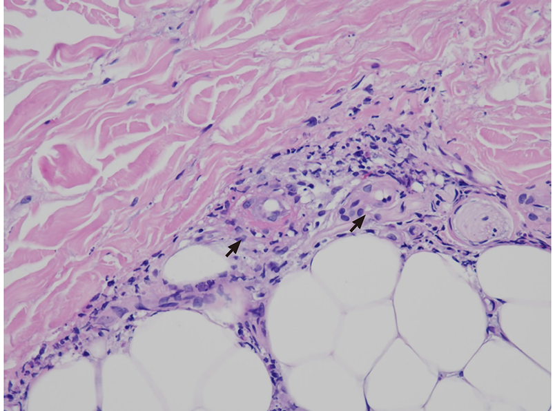
Figure 1 Cryoglobulinemic vasculitis.
The small vessels show neutrophilic inflammation, with fibrinoid necrosis and fragmented neutrophil nuclei (black arrows). Hematoxylin and eosin staining, 400 ×.
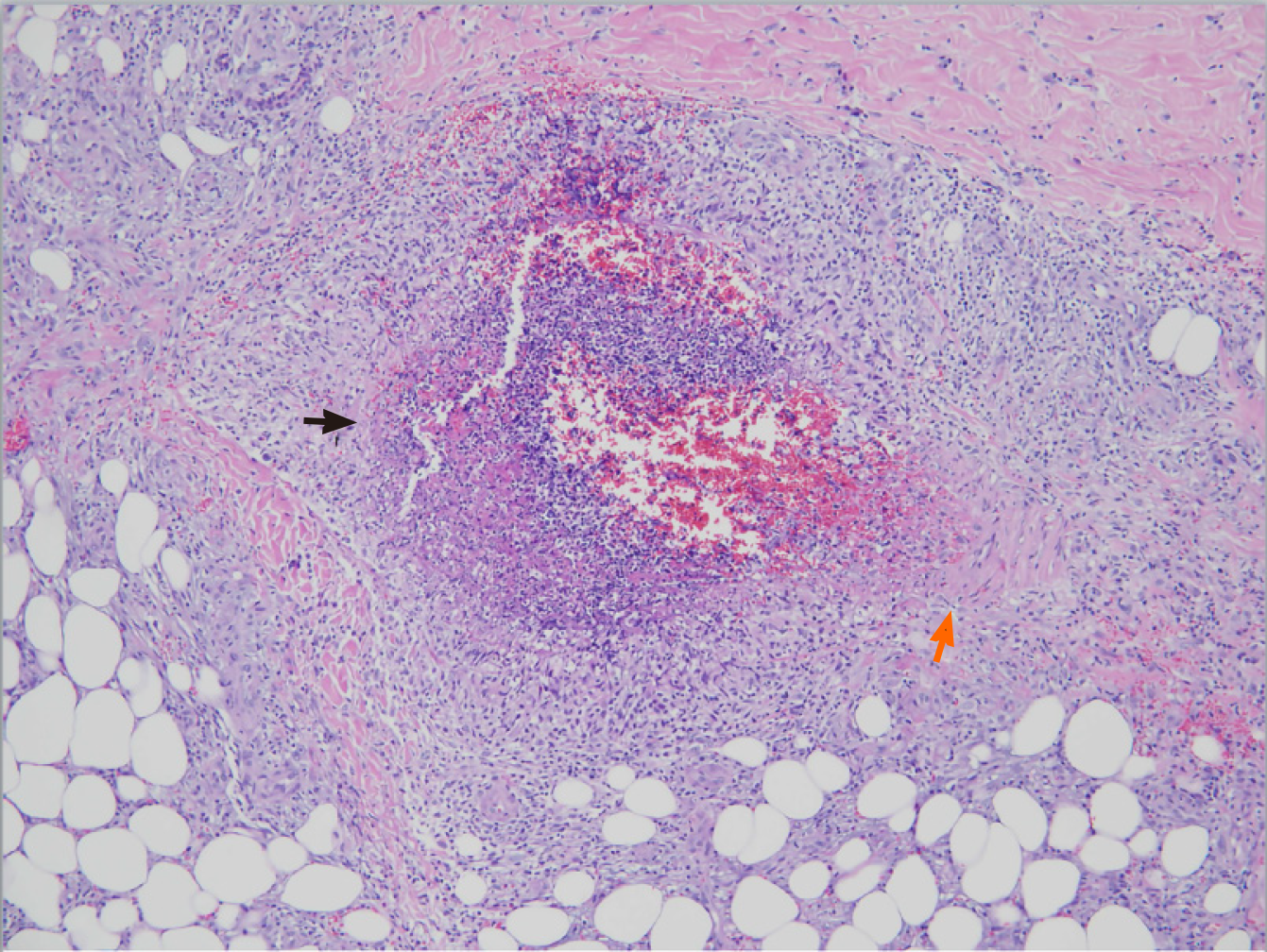
Figure 2 Polyarteritis nodosa.
The vascular wall shows transmural necrotizing inflammation, with intense neutrophilic infiltration and fibrinoid necrosis (black arrow). There is residual muscular wall of the vessel (orange arrow). Hematoxylin and eosin staining, 100 ×.
- Citation: Wang CR, Tsai HW. Human hepatitis viruses-associated cutaneous and systemic vasculitis. World J Gastroenterol 2021; 27(1): 19-36
- URL: https://www.wjgnet.com/1007-9327/full/v27/i1/19.htm
- DOI: https://dx.doi.org/10.3748/wjg.v27.i1.19


